 Movie
Movie Controller

[English] 日本語
 Yorodumi
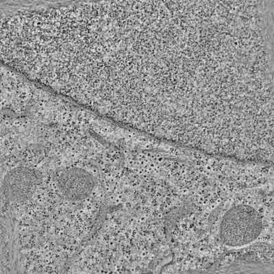
- EMDB-3820: Electron tomographic slices of the nuclear envelope of HeLa cell ... -
+ Open data

- Basic information
| Entry | Database: EMDB / ID: EMD-3820 | |||||||||
|---|---|---|---|---|---|---|---|---|---|---|
| Title | Electron tomographic slices of the nuclear envelope of HeLa cell in interphase | |||||||||
 Map data Map data | None | |||||||||
 Sample Sample |
| |||||||||
| Biological species |  Homo sapiens (human) Homo sapiens (human) | |||||||||
| Method | electron tomography / cryo EM / negative staining | |||||||||
 Authors Authors | Otsuka S / Ellenberg J | |||||||||
 Citation Citation | Journal: Nat Struct Mol Biol / Year: 2018 Title: Postmitotic nuclear pore assembly proceeds by radial dilation of small membrane openings. Authors: Shotaro Otsuka / Anna M Steyer / Martin Schorb / Jean-Karim Hériché / M Julius Hossain / Suruchi Sethi / Moritz Kueblbeck / Yannick Schwab / Martin Beck / Jan Ellenberg /  Abstract: The nuclear envelope has to be reformed after mitosis to create viable daughter cells with closed nuclei. How membrane sealing of DNA and assembly of nuclear pore complexes (NPCs) are achieved and ...The nuclear envelope has to be reformed after mitosis to create viable daughter cells with closed nuclei. How membrane sealing of DNA and assembly of nuclear pore complexes (NPCs) are achieved and coordinated is poorly understood. Here, we reconstructed nuclear membrane topology and the structures of assembling NPCs in a correlative 3D EM time course of dividing human cells. Our quantitative ultrastructural analysis shows that nuclear membranes form from highly fenestrated ER sheets whose holes progressively shrink. NPC precursors are found in small membrane holes and dilate radially during assembly of the inner ring complex, forming thousands of transport channels within minutes. This mechanism is fundamentally different from that of interphase NPC assembly and explains how mitotic cells can rapidly establish a closed nuclear compartment while making it transport competent. | |||||||||
| History |
|
- Structure visualization
| Movie |
 Movie viewer Movie viewer |
|---|---|
| Structure viewer | EM map: SurfViewMolmilJmol/JSmol |
| Supplemental images |
- Downloads & links
-EMDB archive
| Map data | emd_3820.map.gz | 7.5 GB | EMDB map data format | |
|---|---|---|---|---|
| Header (meta data) | emd-3820-v30.xmlemd-3820.xml | 10.7 KB 10.7 KB | Display Display | EMDB header |
| Images |  emd_3820.png emd_3820.png | 139.3 KB | ||
| Archive directory |  http://ftp.pdbj.org/pub/emdb/structures/EMD-3820ftp://ftp.pdbj.org/pub/emdb/structures/EMD-3820 http://ftp.pdbj.org/pub/emdb/structures/EMD-3820ftp://ftp.pdbj.org/pub/emdb/structures/EMD-3820 | HTTPS FTP |
-Related structure data
| EM raw data | EMPIAR-10116 (Title: Raw 2d tomographic tilt series of a dividing cell / Data size: 245.4 Data #1: Raw 2D tilt series for all the electron tomograms [tilt series]) |
|---|
-Links
| EMDB pages | EMDB (EBI/PDBe) / EMDataResource |
|---|
-Map
| File | Download / File: emd_3820.map.gz / Format: CCP4 / Size: 1.8 GB / Type: IMAGE STORED AS SIGNED INTEGER (2 BYTES) | ||||||||||||||||||||||||||||||||||||||||||||||||||||||||||||
|---|---|---|---|---|---|---|---|---|---|---|---|---|---|---|---|---|---|---|---|---|---|---|---|---|---|---|---|---|---|---|---|---|---|---|---|---|---|---|---|---|---|---|---|---|---|---|---|---|---|---|---|---|---|---|---|---|---|---|---|---|---|
| Annotation | None | ||||||||||||||||||||||||||||||||||||||||||||||||||||||||||||
| Projections & slices | Image control
Images are generated by Spider. generated in cubic-lattice coordinate | ||||||||||||||||||||||||||||||||||||||||||||||||||||||||||||
| Voxel size | X=Y=Z: 7.5 Å | ||||||||||||||||||||||||||||||||||||||||||||||||||||||||||||
| Density |
| ||||||||||||||||||||||||||||||||||||||||||||||||||||||||||||
| Symmetry | Space group: 1 | ||||||||||||||||||||||||||||||||||||||||||||||||||||||||||||
| Details | EMDB XML:
CCP4 map header:
| ||||||||||||||||||||||||||||||||||||||||||||||||||||||||||||
 Z (Sec.)
 Y (Row.)
 X (Col.)


-Supplemental data
- Sample components
-Entire : HeLa cell
| Entire | Name: HeLa cell |
|---|---|
| Components |
|
-Supramolecule #1: HeLa cell
| Supramolecule | Name: HeLa cell / type: cell / ID: 1 / Parent: 0 Details: Cells were high-pressure frozen and freeze-substituted into Lowicryl resin. |
|---|---|
| Source (natural) | Organism: Homo sapiens (human) / Strain: HeLa |
-Experimental details
-Structure determination
| Method | negative staining, cryo EM |
|---|---|
 Processing Processing | electron tomography |
| Aggregation state | cell |
-Sample preparation
| Buffer | pH: 7.4 Details: CO2-independent medium without phenol red (Invitrogen), containing 20% FCS, 20% Ficoll PM400 2 mM l-glutamine, and 100 ug/ml penicillin and streptomycin |
|---|---|
| Staining | Type: NEGATIVE / Material: uranyl acetate and lead citrate |
| Sugar embedding | Material: Lowicryl resin Details: Frozen cells were incubated with 0.1% uranyl acetate in acetone at -90C for 20-24 hr and, after infiltration into Lowicryl resin and UV-polymerization, samples were further polymerized by sunlight for 3-4 days. |
| Grid | Model: Grid / Support film - Material: FORMVAR / Support film - topology: CONTINUOUS |
| Vitrification | Cryogen name: NITROGEN |
| High pressure freezing | Instrument: OTHER Details: High pressure freezing chamber was 1.0 mm thick in total, 3.0 mm diameter, with central cavities 50 um deep.. The value given for _emd_high_pressure_freezing.instrument is HPM 010. This is ...Details: High pressure freezing chamber was 1.0 mm thick in total, 3.0 mm diameter, with central cavities 50 um deep.. The value given for _emd_high_pressure_freezing.instrument is HPM 010. This is not in a list of allowed values set(['LEICA EM PACT2', 'LEICA EM PACT', 'EMS-002 RAPID IMMERSION FREEZER', 'OTHER', 'LEICA EM HPM100', 'BAL-TEC HPM 010']) so OTHER is written into the XML file. |
| Cryo protectant | 20% FBS and Ficoll PM400 |
| Sectioning | Ultramicrotomy - Instrument: Leica Ultracut UCT / Ultramicrotomy - Temperature: 25 K / Ultramicrotomy - Final thickness: 300 |
| Fiducial marker | Manufacturer: CMC university Medical Center Utrecht / Diameter: 15 nm |
- Electron microscopy
| Microscope | FEI TECNAI F30 |
|---|---|
| Image recording | Film or detector model: FEI EAGLE (4k x 4k) / Digitization - Dimensions - Width: 4096 pixel / Digitization - Dimensions - Height: 4096 pixel / Number real images: 121 / Average electron dose: 200.0 e/Å2 |
| Electron beam | Acceleration voltage: 300 kV / Electron source:  FIELD EMISSION GUN FIELD EMISSION GUN |
| Electron optics | Illumination mode: FLOOD BEAM / Imaging mode: BRIGHT FIELD / Cs: 2.26 mm / Nominal defocus min: 0.5 µm / Nominal magnification: 15500 |
| Sample stage | Specimen holder model: OTHER |
| Experimental equipment |  Model: Tecnai F30 / Image courtesy: FEI Company |
-Image processing
| Final reconstruction | Algorithm: BACK PROJECTION / Software - Name: IMOD (ver. 4.5.6) Details: Dual axis tilt series were aligned using gold fiducial markers Number images used: 121 |
|---|---|
| CTF correction | Software - Name: IMOD (ver. 4.5.6) |
