 Movie
Movie Controller

[English] 日本語
 Yorodumi
- EMDB-31976: Cryo-EM reconstruction of SARS-CoV-2 M protein dimer in LMNG/CHS ... -
+ Open data

- Basic information
| Entry |  | |||||||||
|---|---|---|---|---|---|---|---|---|---|---|
| Title | Cryo-EM reconstruction of SARS-CoV-2 M protein dimer in LMNG/CHS micelle | |||||||||
 Map data Map data | ||||||||||
 Sample Sample |
| |||||||||
 Keywords Keywords | SARS-CoV-2 / M protein / viral structural protein / virus assembly / VIRAL PROTEIN | |||||||||
| Biological species |   Severe acute respiratory syndrome coronavirus 2 Severe acute respiratory syndrome coronavirus 2 | |||||||||
| Method | single particle reconstruction / cryo EM / Resolution: 6.2 Å | |||||||||
 Authors Authors | Zhang Z / Ohto U / Shimizu T | |||||||||
| Funding support | 1 items
| |||||||||
 Citation Citation | Journal: Nat Commun / Year: 2022 Title: Structure of SARS-CoV-2 membrane protein essential for virus assembly. Authors: Zhikuan Zhang / Norimichi Nomura / Yukiko Muramoto / Toru Ekimoto / Tomoko Uemura / Kehong Liu / Moeko Yui / Nozomu Kono / Junken Aoki / Mitsunori Ikeguchi / Takeshi Noda / So Iwata / ...Authors: Zhikuan Zhang / Norimichi Nomura / Yukiko Muramoto / Toru Ekimoto / Tomoko Uemura / Kehong Liu / Moeko Yui / Nozomu Kono / Junken Aoki / Mitsunori Ikeguchi / Takeshi Noda / So Iwata / Umeharu Ohto / Toshiyuki Shimizu /  Abstract: The coronavirus membrane protein (M) is the most abundant viral structural protein and plays a central role in virus assembly and morphogenesis. However, the process of M protein-driven virus ...The coronavirus membrane protein (M) is the most abundant viral structural protein and plays a central role in virus assembly and morphogenesis. However, the process of M protein-driven virus assembly are largely unknown. Here, we report the cryo-electron microscopy structure of the SARS-CoV-2 M protein in two different conformations. M protein forms a mushroom-shaped dimer, composed of two transmembrane domain-swapped three-helix bundles and two intravirion domains. M protein further assembles into higher-order oligomers. A highly conserved hinge region is key for conformational changes. The M protein dimer is unexpectedly similar to SARS-CoV-2 ORF3a, a viral ion channel. Moreover, the interaction analyses of M protein with nucleocapsid protein (N) and RNA suggest that the M protein mediates the concerted recruitment of these components through the positively charged intravirion domain. Our data shed light on the M protein-driven virus assembly mechanism and provide a structural basis for therapeutic intervention targeting M protein. | |||||||||
| History |
|
- Structure visualization
| Supplemental images |
|---|

- Downloads & links
-EMDB archive
| Map data | emd_31976.map.gz | 7.4 MB |  EMDB map data format EMDB map data format | |
|---|---|---|---|---|
| Header (meta data) | emd-31976-v30.xmlemd-31976.xml | 7.7 KB 7.7 KB | Display Display | EMDB header |
| Images | 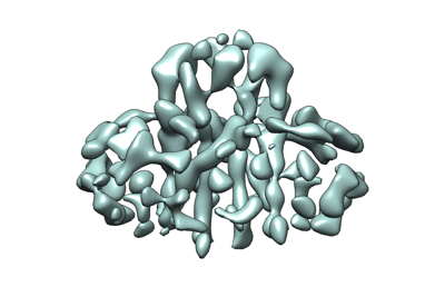 emd_31976.png emd_31976.png | 68.6 KB | ||
| Archive directory |  http://ftp.pdbj.org/pub/emdb/structures/EMD-31976ftp://ftp.pdbj.org/pub/emdb/structures/EMD-31976 http://ftp.pdbj.org/pub/emdb/structures/EMD-31976ftp://ftp.pdbj.org/pub/emdb/structures/EMD-31976 | HTTPS FTP |
-Related structure data
| Related structure data |  7vgrC  7vgsC C: citing same article ( |
|---|---|
| EM raw data | EMPIAR-11168 (Title: Structure of SARS-CoV-2 membrane protein / Data size: 4.4 TB / Data #1: M protein (LMNG/CHS) [micrographs - multiframe] Data #2: M protein (LMNG/CHS) + Fab-E [micrographs - multiframe] Data #3: M protein (LMNG/CHS) + Fab-B [micrographs - multiframe]) |

-Links
| EMDB pages | EMDB (EBI/PDBe) / EMDataResource |
|---|
-Map
| File | Download / File: emd_31976.map.gz / Format: CCP4 / Size: 15.6 MB / Type: IMAGE STORED AS FLOATING POINT NUMBER (4 BYTES) | ||||||||||||||||||||||||||||||||||||
|---|---|---|---|---|---|---|---|---|---|---|---|---|---|---|---|---|---|---|---|---|---|---|---|---|---|---|---|---|---|---|---|---|---|---|---|---|---|
| Projections & slices | Image control
Images are generated by Spider. | ||||||||||||||||||||||||||||||||||||
| Voxel size | X=Y=Z: 1.245 Å | ||||||||||||||||||||||||||||||||||||
| Density |
| ||||||||||||||||||||||||||||||||||||
| Symmetry | Space group: 1 | ||||||||||||||||||||||||||||||||||||
| Details | EMDB XML:
|
 Z (Sec.)
 Y (Row.)
 X (Col.)


-Supplemental data
- Sample components
-Entire : SARS-CoV-2 M protein dimer in LMNG/CHS micelle
| Entire | Name: SARS-CoV-2 M protein dimer in LMNG/CHS micelle |
|---|---|
| Components |
|
-Supramolecule #1: SARS-CoV-2 M protein dimer in LMNG/CHS micelle
| Supramolecule | Name: SARS-CoV-2 M protein dimer in LMNG/CHS micelle / type: complex / ID: 1 / Parent: 0 |
|---|---|
| Source (natural) | Organism: Severe acute respiratory syndrome coronavirus 2 |
-Experimental details
-Structure determination
| Method | cryo EM |
|---|---|
 Processing Processing | single particle reconstruction |
| Aggregation state | particle |
-Sample preparation
| Buffer | pH: 7.5 |
|---|---|
| Vitrification | Cryogen name: ETHANE |
- Electron microscopy
| Microscope | TFS KRIOS |
|---|---|
| Image recording | Film or detector model: GATAN K3 (6k x 4k) / Average electron dose: 57.4 e/Å2 |
| Electron beam | Acceleration voltage: 300 kV / Electron source:  FIELD EMISSION GUN FIELD EMISSION GUN |
| Electron optics | Illumination mode: OTHER / Imaging mode: BRIGHT FIELD |
| Experimental equipment |  Model: Titan Krios / Image courtesy: FEI Company |
-Image processing
| Startup model | Type of model: OTHER |
|---|---|
| Final reconstruction | Applied symmetry - Point group: C2 (2 fold cyclic) / Resolution.type: BY AUTHOR / Resolution: 6.2 Å / Resolution method: FSC 0.143 CUT-OFF / Number images used: 348142 |
| Initial angle assignment | Type: RANDOM ASSIGNMENT |
| Final angle assignment | Type: RANDOM ASSIGNMENT |
