 Movie
Movie Controller

[English] 日本語
 Yorodumi
- EMDB-29393: Mouse apoferritin from data collected on Titan Krios with K3 came... -
+ Open data

- Basic information
| Entry |  | |||||||||
|---|---|---|---|---|---|---|---|---|---|---|
| Title | Mouse apoferritin from data collected on Titan Krios with K3 camera in counting mode with a 20 eV energy filter slit, in 200-500 nm Ice thickness | |||||||||
 Map data Map data | Mouse apoferritin from data collected on Titan Krios with K3 camera in counting mode in 200-500 nm Ice thickness | |||||||||
 Sample Sample |
| |||||||||
 Keywords Keywords | Mouse apoferritin / Titan Krios / K3 camera / counting mode / 200-500 nm ice thickness / METAL BINDING PROTEIN | |||||||||
| Biological species |  | |||||||||
| Method | single particle reconstruction / cryo EM / Resolution: 2.9 Å | |||||||||
 Authors Authors | Neselu K / Wang B / Rice WJ / Potter CS / Carragher B / Chua EYD | |||||||||
| Funding support |  United States, 2 items United States, 2 items
| |||||||||
 Citation Citation | Journal: J Struct Biol X / Year: 2023 Title: Measuring the effects of ice thickness on resolution in single particle cryo-EM. Authors: Kasahun Neselu / Bing Wang / William J Rice / Clinton S Potter / Bridget Carragher / Eugene Y D Chua / Abstract: Ice thickness is a critical parameter in single particle cryo-EM - too thin ice can break during imaging or exclude the sample of interest, while ice that is too thick contributes to more inelastic ...Ice thickness is a critical parameter in single particle cryo-EM - too thin ice can break during imaging or exclude the sample of interest, while ice that is too thick contributes to more inelastic scattering that precludes obtaining high resolution reconstructions. Here we present the practical effects of ice thickness on resolution, and the influence of energy filters, accelerating voltage, or detector mode. We collected apoferritin data with a wide range of ice thicknesses on three microscopes with different instrumentation and settings. We show that on a 300 kV microscope, using a 20 eV energy filter slit has a greater effect on improving resolution in thicker ice; that operating at 300 kV instead of 200 kV accelerating voltage provides significant resolution improvements at an ice thickness above 150 nm; and that on a 200 kV microscope using a detector operating in super resolution mode enables good reconstructions for up to 200 nm ice thickness, while collecting in counting instead of linear mode leads to improvements in resolution for ice of 50-150 nm thickness. Our findings can serve as a guide for users seeking to optimize data collection or sample preparation routines for both single particle and in situ cryo-EM. We note that most in situ data collection is done on samples in a range of ice thickness above 150 nm so these results may be especially relevant to that community. | |||||||||
| History |
|
- Structure visualization
| Supplemental images |
|---|

- Downloads & links
-EMDB archive
| Map data | emd_29393.map.gz | 59.6 MB |  EMDB map data format EMDB map data format | |
|---|---|---|---|---|
| Header (meta data) | emd-29393-v30.xmlemd-29393.xml | 15.2 KB 15.2 KB | Display Display | EMDB header |
| FSC (resolution estimation) | emd_29393_fsc.xml | 8.4 KB | Display | FSC data file |
| Images | 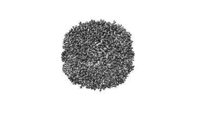 emd_29393.png emd_29393.png | 39.4 KB | ||
| Filedesc metadata | emd-29393.cif.gz | 4.4 KB | ||
| Others | emd_29393_half_map_1.map.gzemd_29393_half_map_2.map.gz | 59 MB 59 MB | ||
| Archive directory |  http://ftp.pdbj.org/pub/emdb/structures/EMD-29393ftp://ftp.pdbj.org/pub/emdb/structures/EMD-29393 http://ftp.pdbj.org/pub/emdb/structures/EMD-29393ftp://ftp.pdbj.org/pub/emdb/structures/EMD-29393 | HTTPS FTP |
-Related structure data
| Related structure data | C: citing same article ( |
|---|


-Links
| EMDB pages | EMDB (EBI/PDBe) / EMDataResource |
|---|
-Map
| File | Download / File: emd_29393.map.gz / Format: CCP4 / Size: 64 MB / Type: IMAGE STORED AS FLOATING POINT NUMBER (4 BYTES) | ||||||||||||||||||||||||||||||||||||
|---|---|---|---|---|---|---|---|---|---|---|---|---|---|---|---|---|---|---|---|---|---|---|---|---|---|---|---|---|---|---|---|---|---|---|---|---|---|
| Annotation | Mouse apoferritin from data collected on Titan Krios with K3 camera in counting mode in 200-500 nm Ice thickness | ||||||||||||||||||||||||||||||||||||
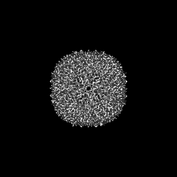
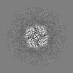
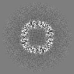
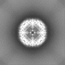
| Projections & slices | Image control
Images are generated by Spider. | ||||||||||||||||||||||||||||||||||||
| Voxel size | X=Y=Z: 1.083 Å | ||||||||||||||||||||||||||||||||||||
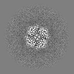
| Density |
| ||||||||||||||||||||||||||||||||||||
| Symmetry | Space group: 1 | ||||||||||||||||||||||||||||||||||||
| Details | EMDB XML:
|
 Z (Sec.)
 Y (Row.)
 X (Col.)


-Supplemental data
-Half map: Mouse apoferritin from data collected on Titan Krios...
| File | emd_29393_half_map_1.map | ||||||||||||
|---|---|---|---|---|---|---|---|---|---|---|---|---|---|
| Annotation | Mouse apoferritin from data collected on Titan Krios with K3 camera in counting mode in 200-500 nm Ice thickness | ||||||||||||
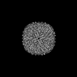
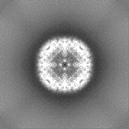
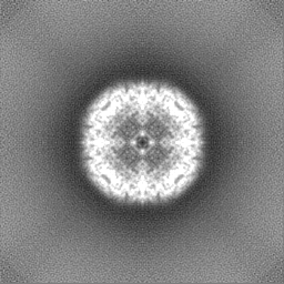
| Projections & Slices |
| ||||||||||||
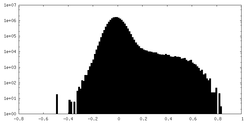
| Density Histograms |


-Half map: Mouse apoferritin from data collected on Titan Krios...
| File | emd_29393_half_map_2.map | ||||||||||||
|---|---|---|---|---|---|---|---|---|---|---|---|---|---|
| Annotation | Mouse apoferritin from data collected on Titan Krios with K3 camera in counting mode in 200-500 nm Ice thickness | ||||||||||||
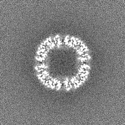
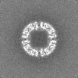
| Projections & Slices |
| ||||||||||||
| Density Histograms |


- Sample components
-Entire : Mouse apoferritin
| Entire | Name: Mouse apoferritin |
|---|---|
| Components |
|
-Supramolecule #1: Mouse apoferritin
| Supramolecule | Name: Mouse apoferritin / type: complex / ID: 1 / Parent: 0 |
|---|---|
| Source (natural) | Organism: |
| Molecular weight | Theoretical: 450 KDa |
-Experimental details
-Structure determination
| Method | cryo EM |
|---|---|
 Processing Processing | single particle reconstruction |
| Aggregation state | particle |
-Sample preparation
| Concentration | 8 mg/mL | ||||||||||||
|---|---|---|---|---|---|---|---|---|---|---|---|---|---|
| Buffer | pH: 7.5 Component:
| ||||||||||||
| Grid | Model: UltrAuFoil R1.2/1.3 / Material: GOLD / Mesh: 300 / Support film - Material: GOLD / Support film - topology: HOLEY / Pretreatment - Type: PLASMA CLEANING / Pretreatment - Time: 10 sec. / Pretreatment - Atmosphere: OTHER | ||||||||||||
| Vitrification | Cryogen name: ETHANE / Chamber humidity: 100 % / Chamber temperature: 295.15 K / Instrument: FEI VITROBOT MARK IV |
- Electron microscopy
| Microscope | FEI TITAN KRIOS |
|---|---|
| Specialist optics | Energy filter - Name: GIF Bioquantum / Energy filter - Slit width: 20 eV |
| Image recording | Film or detector model: GATAN K3 BIOQUANTUM (6k x 4k) / Number grids imaged: 1 / Average exposure time: 2.0 sec. / Average electron dose: 51.22 e/Å2 |
| Electron beam | Acceleration voltage: 300 kV / Electron source:  FIELD EMISSION GUN FIELD EMISSION GUN |
| Electron optics | C2 aperture diameter: 100.0 µm / Illumination mode: FLOOD BEAM / Imaging mode: BRIGHT FIELD / Cs: 2.7 mm / Nominal defocus max: 2.0 µm / Nominal defocus min: 0.8 µm / Nominal magnification: 81000 |
| Sample stage | Specimen holder model: FEI TITAN KRIOS AUTOGRID HOLDER |
| Experimental equipment |  Model: Titan Krios / Image courtesy: FEI Company |

