HslUV protease complex / endopeptidase Clp / endopeptidase Clp complex / positive regulation of programmed cell death / response to temperature stimulus / ATP-dependent peptidase activity / protein quality control for misfolded or incompletely synthesized proteins / proteasomal protein catabolic process / serine-type peptidase activity / : ...HslUV protease complex / endopeptidase Clp / endopeptidase Clp complex / positive regulation of programmed cell death / response to temperature stimulus / ATP-dependent peptidase activity / protein quality control for misfolded or incompletely synthesized proteins / proteasomal protein catabolic process / serine-type peptidase activity / : / bioluminescence / generation of precursor metabolites and energy / ATP-dependent protein folding chaperone / response to radiation / positive regulation of protein catabolic process / : / peptidase activity / ATPase binding / response to heat / protein dimerization activity / ribosome / serine-type endopeptidase activity / cell division / ATP hydrolysis activity / zinc ion binding / ATP binding / membrane / identical protein binding / cytosol Similarity search - Function
Stringent starvation protein B / SspB-like superfamily / Stringent starvation protein B / Zinc finger, ClpX C4-type superfamily / ClpX C4-type zinc finger / Zinc finger, ClpX C4-type / Clp protease, ATP-binding subunit ClpX, bacteria / ClpX C4-type zinc finger / : / Clp protease, ATP-binding subunit ClpX ...Stringent starvation protein B / SspB-like superfamily / Stringent starvation protein B / Zinc finger, ClpX C4-type superfamily / ClpX C4-type zinc finger / Zinc finger, ClpX C4-type / Clp protease, ATP-binding subunit ClpX, bacteria / ClpX C4-type zinc finger / : / Clp protease, ATP-binding subunit ClpX / ClpX zinc binding (ZB) domain profile. / : / ClpP, Ser active site / Endopeptidase Clp serine active site. / ClpP, histidine active site / Endopeptidase Clp histidine active site. / ATP-dependent Clp protease proteolytic subunit / Clp protease proteolytic subunit /Translocation-enhancing protein TepA / Clp protease / Clp ATPase, C-terminal / C-terminal, D2-small domain, of ClpB protein / C-terminal, D2-small domain, of ClpB protein / AAA domain (Cdc48 subfamily) / ClpP/crotonase-like domain superfamily / Green fluorescent protein, GFP / Green fluorescent protein-related / Green fluorescent protein / Green fluorescent protein / ATPase, AAA-type, core / ATPases associated with a variety of cellular activities / AAA+ ATPase domain / P-loop containing nucleoside triphosphate hydrolase Similarity search - Domain/homology
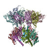
ATP-dependent Clp protease ATP-binding subunit ClpX / ATP-dependent Clp protease proteolytic subunit / Stringent starvation protein B / Green fluorescent protein Similarity search - Component
Biological species
Escherichia coli (E. coli)
Method
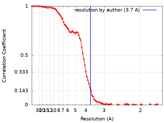
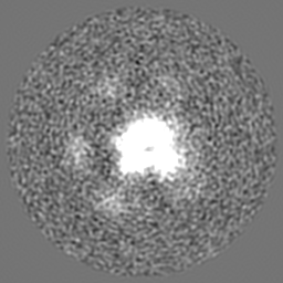
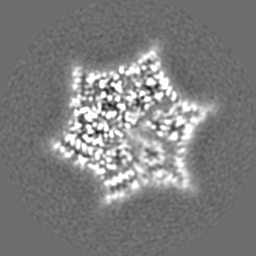
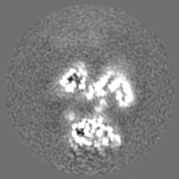
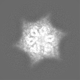
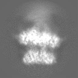
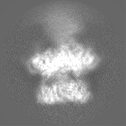
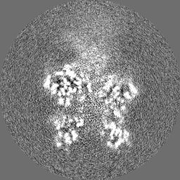
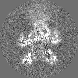
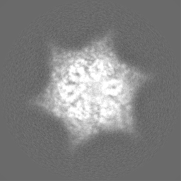
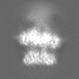
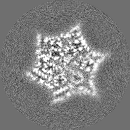
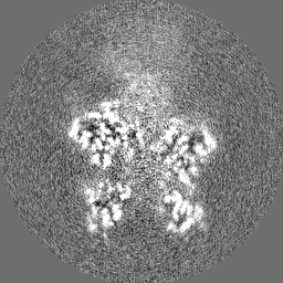
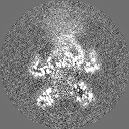
single particle reconstruction / cryo EM / Resolution: 3.7 Å
National Institutes of Health/National Institute of General Medical Sciences (NIH/NIGMS)
United States
Citation
Journal: Proc Natl Acad Sci U S A / Year: 2023 Title: The SspB adaptor drives structural changes in the AAA+ ClpXP protease during ssrA-tagged substrate delivery. Authors: Alireza Ghanbarpour / Xue Fei / Tania A Baker / Joseph H Davis / Robert T Sauer / Abstract: Energy-dependent protein degradation by the AAA+ ClpXP protease helps maintain protein homeostasis in bacteria and eukaryotic organelles of bacterial origin. In and many other proteobacteria, the ...Energy-dependent protein degradation by the AAA+ ClpXP protease helps maintain protein homeostasis in bacteria and eukaryotic organelles of bacterial origin. In and many other proteobacteria, the SspB adaptor assists ClpXP in degrading ssrA-tagged polypeptides produced as a consequence of tmRNA-mediated ribosome rescue. By tethering these incomplete ssrA-tagged proteins to ClpXP, SspB facilitates their efficient degradation at low substrate concentrations. How this process occurs structurally is unknown. Here, we present a cryo-EM structure of the SspB adaptor bound to a GFP-ssrA substrate and to ClpXP. This structure provides evidence for simultaneous contacts of SspB and ClpX with the ssrA tag within the tethering complex, allowing direct substrate handoff concomitant with the initiation of substrate translocation. Furthermore, our structure reveals that binding of the substrate·adaptor complex induces unexpected conformational changes within the spiral structure of the AAA+ ClpX hexamer and its interaction with the ClpP tetradecamer.
In the structure databanks used in Yorodumi, some data are registered as the other names, "COVID-19 virus" and "2019-nCoV". Here are the details of the virus and the list of structure data.
Jan 31, 2019. EMDB accession codes are about to change! (news from PDBe EMDB page)
EMDB accession codes are about to change! (news from PDBe EMDB page)
The allocation of 4 digits for EMDB accession codes will soon come to an end. Whilst these codes will remain in use, new EMDB accession codes will include an additional digit and will expand incrementally as the available range of codes is exhausted. The current 4-digit format prefixed with “EMD-” (i.e. EMD-XXXX) will advance to a 5-digit format (i.e. EMD-XXXXX), and so on. It is currently estimated that the 4-digit codes will be depleted around Spring 2019, at which point the 5-digit format will come into force.
The EM Navigator/Yorodumi systems omit the EMD- prefix.
Related info.:Q: What is EMD? / ID/Accession-code notation in Yorodumi/EM Navigator
Yorodumi is a browser for structure data from EMDB, PDB, SASBDB, etc.
This page is also the successor to EM Navigator detail page, and also detail information page/front-end page for Omokage search.
The word "yorodu" (or yorozu) is an old Japanese word meaning "ten thousand". "mi" (miru) is to see.
Related info.:EMDB / PDB / SASBDB / Comparison of 3 databanks / Yorodumi Search / Aug 31, 2016. New EM Navigator & Yorodumi / Yorodumi Papers / Jmol/JSmol / Function and homology information / Changes in new EM Navigator and Yorodumi
 Movie
Movie Controller

 Yorodumi
 Open data
 Basic information
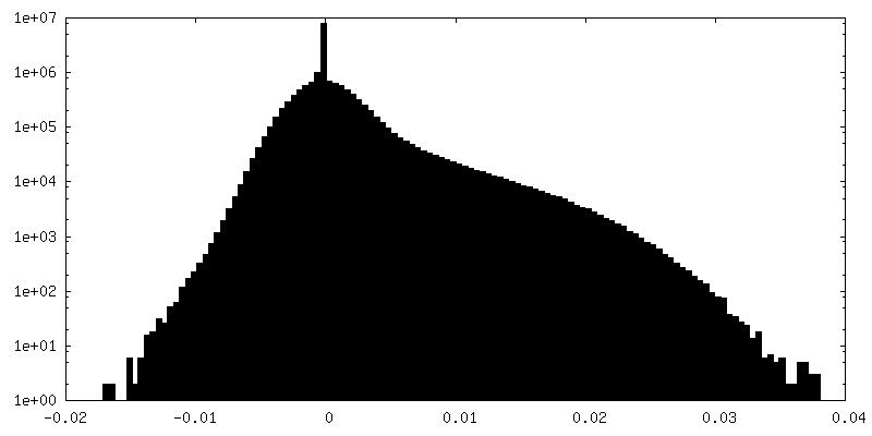
 Map data
 Sample
 Keywords
 Function and homology information
 Authors
 United States, 1 items
  Citation
 Structure visualization
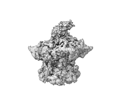
 Downloads & links
 emd_28585.png
 http://ftp.pdbj.org/pub/emdb/structures/EMD-28585
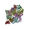


 Z (Sec.)
 Y (Row.)
 X (Col.)


 Sample components

 Processing
 Electron microscopy
 FIELD EMISSION GUN