 Movie
Movie Controller


+ Open data

- Basic information
| Entry |  | |||||||||
|---|---|---|---|---|---|---|---|---|---|---|
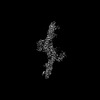
| Title | Gea2 closed/open conformation (composite structure) | |||||||||
 Map data Map data | Gea2 closed/open conformation (composite structure) | |||||||||
 Sample Sample |
| |||||||||
 Keywords Keywords | GEF / PROTEIN TRANSPORT | |||||||||
| Function / homology |  Function and homology information Function and homology informationcellular localization / regulation of ARF protein signal transduction / vesicle-mediated transport / guanyl-nucleotide exchange factor activity / Golgi apparatus Similarity search - Function | |||||||||
| Biological species |  | |||||||||
| Method | single particle reconstruction / cryo EM / Resolution: 4.7 Å | |||||||||
 Authors Authors | Muccini A / Fromme JC | |||||||||
| Funding support |  United States, 1 items United States, 1 items
| |||||||||
 Citation Citation | Journal: Cell Rep / Year: 2022 Title: Structural basis for activation of Arf1 at the Golgi complex. Authors: Arnold J Muccini / Margaret A Gustafson / J Christopher Fromme / Abstract: The Golgi complex is the central sorting station of the eukaryotic secretory pathway. Traffic through the Golgi requires activation of Arf guanosine triphosphatases that orchestrate cargo sorting and ...The Golgi complex is the central sorting station of the eukaryotic secretory pathway. Traffic through the Golgi requires activation of Arf guanosine triphosphatases that orchestrate cargo sorting and vesicle formation by recruiting an array of effector proteins. Arf activation and Golgi membrane association is controlled by large guanine nucleotide exchange factors (GEFs) possessing multiple conserved regulatory domains. Here we present cryoelectron microscopy (cryoEM) structures of full-length Gea2, the yeast paralog of the human Arf-GEF GBF1, that reveal the organization of these regulatory domains and explain how Gea2 binds to the Golgi membrane surface. We find that the GEF domain adopts two different conformations compatible with different stages of the Arf activation reaction. The structure of a Gea2-Arf1 activation intermediate suggests that the movement of the GEF domain primes Arf1 for membrane insertion upon guanosine triphosphate binding. We propose that conformational switching of Gea2 during the nucleotide exchange reaction promotes membrane insertion of Arf1. | |||||||||
| History |
|
- Structure visualization
| Supplemental images |
|---|

- Downloads & links
-EMDB archive
| Map data | emd_26717.map.gz | 81.3 MB | EMDB map data format | |
|---|---|---|---|---|
| Header (meta data) | emd-26717-v30.xmlemd-26717.xml | 9.9 KB 9.9 KB | Display Display | EMDB header |
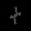
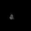
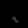
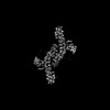
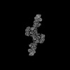
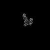
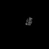
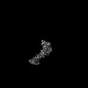
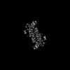
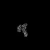
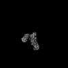
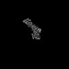
| Images | 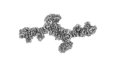 emd_26717.png emd_26717.png | 34.5 KB | ||
| Filedesc metadata | emd-26717.cif.gz | 5.7 KB | ||
| Archive directory |  http://ftp.pdbj.org/pub/emdb/structures/EMD-26717ftp://ftp.pdbj.org/pub/emdb/structures/EMD-26717 http://ftp.pdbj.org/pub/emdb/structures/EMD-26717ftp://ftp.pdbj.org/pub/emdb/structures/EMD-26717 | HTTPS FTP |
-Related structure data
| Related structure data | 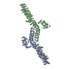 7urrMC  7uroC 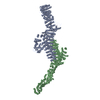 7ut4C  7uthC M: atomic model generated by this map C: citing same article ( |
|---|---|
| Similar structure data |


-Links
| EMDB pages | EMDB (EBI/PDBe) / EMDataResource |
|---|
-Map
| File | Download / File: emd_26717.map.gz / Format: CCP4 / Size: 103 MB / Type: IMAGE STORED AS FLOATING POINT NUMBER (4 BYTES) | ||||||||||||||||||||||||||||||||||||
|---|---|---|---|---|---|---|---|---|---|---|---|---|---|---|---|---|---|---|---|---|---|---|---|---|---|---|---|---|---|---|---|---|---|---|---|---|---|
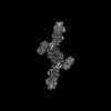
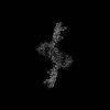
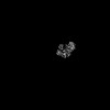
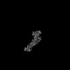
| Annotation | Gea2 closed/open conformation (composite structure) | ||||||||||||||||||||||||||||||||||||
| Projections & slices | Image control
Images are generated by Spider. | ||||||||||||||||||||||||||||||||||||
| Voxel size | X=Y=Z: 1.664 Å | ||||||||||||||||||||||||||||||||||||
| Density |
| ||||||||||||||||||||||||||||||||||||
| Symmetry | Space group: 1 | ||||||||||||||||||||||||||||||||||||
| Details | EMDB XML:
|
 X (Sec.)
 Y (Row.)
 Z (Col.)


-Supplemental data
- Sample components
-Entire : Gea2
| Entire | Name: Gea2 |
|---|---|
| Components |
|
-Supramolecule #1: Gea2
| Supramolecule | Name: Gea2 / type: complex / ID: 1 / Parent: 0 / Macromolecule list: all |
|---|---|
| Source (natural) | Organism: |
-Macromolecule #1: GEA2 isoform 1
| Macromolecule | Name: GEA2 isoform 1 / type: protein_or_peptide / ID: 1 / Number of copies: 2 / Enantiomer: LEVO |
|---|---|
| Source (natural) | Organism: |
| Molecular weight | Theoretical: 165.846844 KDa |
| Recombinant expression | Organism:  Komagataella pastoris (fungus) Komagataella pastoris (fungus) |
| Sequence | String: MSDREFVTVD PVTIIIKECI NLSTAMRKYS KFTSQSGVAA LLGGGSEIFS NQDDYLAHTF NNLNTNKHND PFLSGFIQLR LMLNKLKNL DNIDSLTILQ PFLLIVSTSS ISGYITSLAL DSLQKFFTLN IINESSQNYI GAHRATVNAL THCRFEGSQQ L SDDSVLLK ...String: MSDREFVTVD PVTIIIKECI NLSTAMRKYS KFTSQSGVAA LLGGGSEIFS NQDDYLAHTF NNLNTNKHND PFLSGFIQLR LMLNKLKNL DNIDSLTILQ PFLLIVSTSS ISGYITSLAL DSLQKFFTLN IINESSQNYI GAHRATVNAL THCRFEGSQQ L SDDSVLLK VVFLLRSIVD SPYGDLLSNS IIYDVLQTIL SLACNNRRSE VLRNAAQSTM IAVTVKIFSK LKTIEPVNVN QI YINDESY TNDVLKADTI GTNVESKEEG SQEDPIGMKV NNEEAISEDD GIEEEHIHSE KSTNGAEQLD IVQKTTRSNS RIQ AYADDN YGLPVVRQYL NLLLSLIAPE NELKHSYSTR IFGLELIQTA LEISGDRLQL YPRLFTLISD PIFKSILFII QNTT KLSLL QATLQLFTTL VVILGNNLQL QIELTLTRIF SILLDDGTAN NSSSENKNKP SIIKELLIEQ ISILWTRSPS FFTST FINF DCNLDRADVS INFLKALTKL ALPESALTTT ESVPPICLEG LVSLVDDMFD HMKDIDREEF GRQKNEMEIL KKRDRK TEF IECTNAFNEK PKKGIPMLIE KGFIASDSDK DIAEFLFNNN NRMNKKTIGL LLCHPDKVSL LNEYIRLFDF SGLRVDE AI RILLTKFRLP GESQQIERII EAFSSAYCEN QDYDPSKISD NAEDDISTVQ PDADSVFILS YSIIMLNTDL HNPQVKEH M SFEDYSGNLK GCCNHKDFPF WYLDRIYCSI RDKEIVMPEE HHGNEKWFED AWNNLISSTT VITEIKKDTQ SVMDKLTPL ELLNFDRAIF KQVGPSIVST LFNIYVVASD DHISTRMITS LDKCSYISAF FDFKDLFNDI LNSIAKGTTL INSSHDDELS TLAFEYGPM PLVQIKFEDT NTEIPVSTDA VRFGRSFKGQ LNTVVFFRII RRNKDPKIFS KELWLNIVNI ILTLYEDLIL S PDIFPDLQ KRLKLSNLPK PSPEISINKS KESKGLLSTF ASYLKGDEEP TEEEIKSSKK AMECIKSSNI AASVFGNESN IT ADLIKTL LDSAKTEKNA DNSRYFEAEL LFIIELTIAL FLFCKEEKEL GKFILQKVFQ LSHTKGLTKR TVRRMLTYKI LLI SLCADQ TEYLSKLIND ELLKKGDIFT QKFFATNQGK EFLKRLFSLT ESEFYRGFLL GNENFWKFLR KVTAMKEQSE SIFE YLNES IKTDSNILTN ENFMWVLGLL DEISSMGAVG NHWEIEYKKL TESGHKIDKE NPYKKSIELS LKSIQLTSHL LEDNN DLRK NEIFAIIQAL AHQCINPCKQ ISEFAVVTLE QTLINKIEIP TNEMESVEEL IEGGLLPLLN SSETQEDQKI LISSIL TII SNVYLHYLKL GKTSNETFLK ILSIFNKFVE DSDIEKKLQQ LILDKKSIEK GNGSSSHGSA HEQTPESNDV EIEATAP ID DNTDDDNKPK LSDVEKD UniProtKB: GEA2 isoform 1 |
-Experimental details
-Structure determination
| Method | cryo EM |
|---|---|
 Processing Processing | single particle reconstruction |
| Aggregation state | particle |
-Sample preparation
| Buffer | pH: 8 |
|---|---|
| Vitrification | Cryogen name: ETHANE |
- Electron microscopy
| Microscope | FEI TALOS ARCTICA |
|---|---|
| Image recording | Film or detector model: GATAN K3 (6k x 4k) / Average electron dose: 1.0 e/Å2 |
| Electron beam | Acceleration voltage: 200 kV / Electron source:  FIELD EMISSION GUN FIELD EMISSION GUN |
| Electron optics | Illumination mode: FLOOD BEAM / Imaging mode: BRIGHT FIELD / Nominal defocus max: 2.0 µm / Nominal defocus min: 1.0 µm |
| Experimental equipment |  Model: Talos Arctica / Image courtesy: FEI Company |
-Image processing
| Startup model | Type of model: INSILICO MODEL |
|---|---|
| Final reconstruction | Resolution.type: BY AUTHOR / Resolution: 4.7 Å / Resolution method: FSC 0.143 CUT-OFF / Number images used: 101014 |
| Initial angle assignment | Type: MAXIMUM LIKELIHOOD |
| Final angle assignment | Type: MAXIMUM LIKELIHOOD |
