 Movie
Movie Controller

[English] 日本語
 Yorodumi
- EMDB-26434: Structure of RecT protein from Listeria innoccua phage A118 in co... -
+ Open data

- Basic information
| Entry | 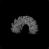 | |||||||||
|---|---|---|---|---|---|---|---|---|---|---|
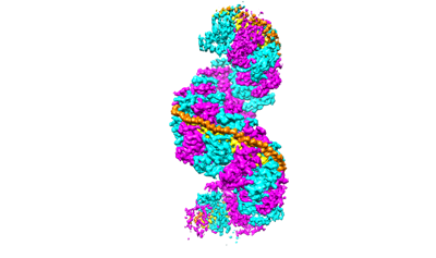
| Title | Structure of RecT protein from Listeria innoccua phage A118 in complex with 83-mer annealed duplex | |||||||||
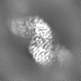
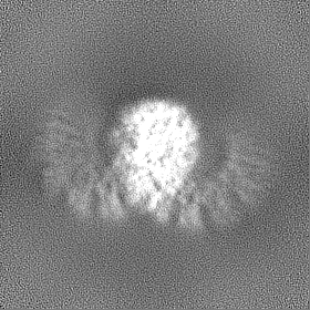
 Map data Map data | Density modified map from Phenix-Resolve, including 10-fold NCS averaging. | |||||||||
 Sample Sample |
| |||||||||
 Keywords Keywords | DNA Recombination / DNA Annealing / DNA BINDING PROTEIN / DNA BINDING PROTEIN-DNA complex | |||||||||
| Function / homology | DNA single-strand annealing protein RecT / RecT family / RecT family / DNA metabolic process / DNA binding / Recombinase [Bacteriophage A118] Function and homology information Function and homology information | |||||||||
| Biological species |  Listeria innocua Clip11262 (bacteria) / Listeria innocua Clip11262 (bacteria) /  Escherichia virus M13 Escherichia virus M13 | |||||||||
| Method | single particle reconstruction / cryo EM / Resolution: 3.4 Å | |||||||||
 Authors Authors | Bell CE / Caldwell BJ | |||||||||
| Funding support |  United States, 1 items United States, 1 items
| |||||||||
 Citation Citation | Journal: Nat Commun / Year: 2022 Title: Structure of a RecT/Redβ family recombinase in complex with a duplex intermediate of DNA annealing. Authors: Brian J Caldwell / Andrew S Norris / Caroline F Karbowski / Alyssa M Wiegand / Vicki H Wysocki / Charles E Bell / Abstract: Some bacteriophage encode a recombinase that catalyzes single-stranded DNA annealing (SSA). These proteins are apparently related to RAD52, the primary human SSA protein. The best studied protein, ...Some bacteriophage encode a recombinase that catalyzes single-stranded DNA annealing (SSA). These proteins are apparently related to RAD52, the primary human SSA protein. The best studied protein, Redβ from bacteriophage λ, binds weakly to ssDNA, not at all to dsDNA, but tightly to a duplex intermediate of annealing formed when two complementary DNA strands are added to the protein sequentially. We used single particle cryo-electron microscopy (cryo-EM) to determine a 3.4 Å structure of a Redβ homolog from a prophage of Listeria innocua in complex with two complementary 83mer oligonucleotides. The structure reveals a helical protein filament bound to a DNA duplex that is highly extended and unwound. Native mass spectrometry confirms that the complex seen by cryo-EM is the predominant species in solution. The protein shares a common core fold with RAD52 and a similar mode of ssDNA-binding. These data provide insights into the mechanism of protein-catalyzed SSA. | |||||||||
| History |
|
- Structure visualization
| Supplemental images |
|---|
- Downloads & links
-EMDB archive
| Map data | emd_26434.map.gz | 77.9 MB | EMDB map data format | |
|---|---|---|---|---|
| Header (meta data) | emd-26434-v30.xmlemd-26434.xml | 19.8 KB 19.8 KB | Display Display | EMDB header |
| FSC (resolution estimation) | emd_26434_fsc.xml | 7.8 KB | Display | FSC data file |
| Images |  emd_26434.png emd_26434.png | 46.5 KB | ||
| Filedesc metadata | emd-26434.cif.gz | 6.7 KB | ||
| Others | emd_26434_half_map_1.map.gzemd_26434_half_map_2.map.gz | 77.6 MB 77.6 MB | ||
| Archive directory |  http://ftp.pdbj.org/pub/emdb/structures/EMD-26434ftp://ftp.pdbj.org/pub/emdb/structures/EMD-26434 http://ftp.pdbj.org/pub/emdb/structures/EMD-26434ftp://ftp.pdbj.org/pub/emdb/structures/EMD-26434 | HTTPS FTP |
-Related structure data
| Related structure data |  7ub2MC  7ubbC M: atomic model generated by this map C: citing same article ( |
|---|---|
| Similar structure data |

-Links
| EMDB pages | EMDB (EBI/PDBe) / EMDataResource |
|---|
-Map
| File | Download / File: emd_26434.map.gz / Format: CCP4 / Size: 83.7 MB / Type: IMAGE STORED AS FLOATING POINT NUMBER (4 BYTES) | ||||||||||||||||||||||||||||||||||||
|---|---|---|---|---|---|---|---|---|---|---|---|---|---|---|---|---|---|---|---|---|---|---|---|---|---|---|---|---|---|---|---|---|---|---|---|---|---|
| Annotation | Density modified map from Phenix-Resolve, including 10-fold NCS averaging. | ||||||||||||||||||||||||||||||||||||
| Projections & slices | Image control
Images are generated by Spider. | ||||||||||||||||||||||||||||||||||||
| Voxel size | X=Y=Z: 0.899 Å | ||||||||||||||||||||||||||||||||||||
| Density |
| ||||||||||||||||||||||||||||||||||||
| Symmetry | Space group: 1 | ||||||||||||||||||||||||||||||||||||
| Details | EMDB XML:
|
 X (Sec.)
 Y (Row.)
 Z (Col.)


-Supplemental data
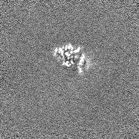
-Half map: Un-masked half map
| File | emd_26434_half_map_1.map | ||||||||||||
|---|---|---|---|---|---|---|---|---|---|---|---|---|---|
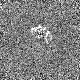
| Annotation | Un-masked half map | ||||||||||||
| Projections & Slices |
| ||||||||||||
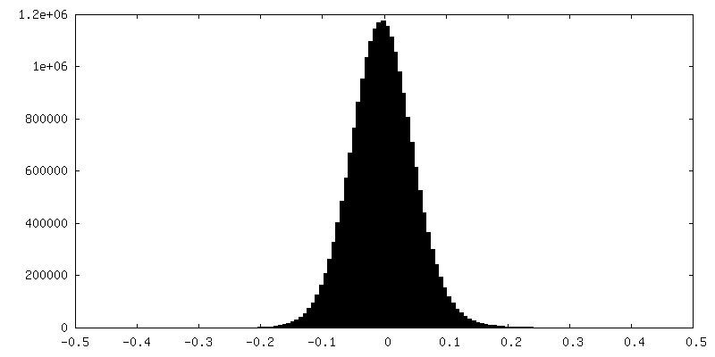
| Density Histograms |


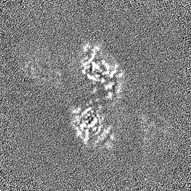
-Half map: Un-masked half map
| File | emd_26434_half_map_2.map | ||||||||||||
|---|---|---|---|---|---|---|---|---|---|---|---|---|---|
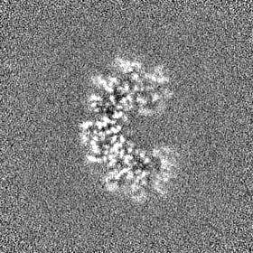
| Annotation | Un-masked half map | ||||||||||||
| Projections & Slices |
| ||||||||||||
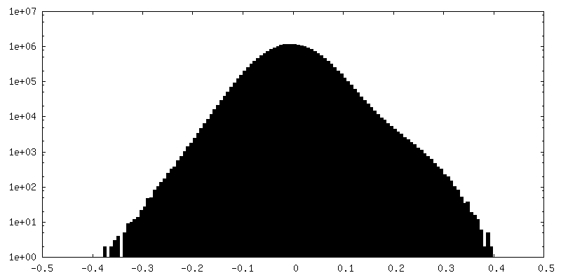
| Density Histograms |


- Sample components
-Entire : RecT protein from Listeria innocua phage A118, complexed with two...
| Entire | Name: RecT protein from Listeria innocua phage A118, complexed with two complementary strands of ssDNA that were added to the protein sequentially |
|---|---|
| Components |
|
-Supramolecule #1: RecT protein from Listeria innocua phage A118, complexed with two...
| Supramolecule | Name: RecT protein from Listeria innocua phage A118, complexed with two complementary strands of ssDNA that were added to the protein sequentially type: complex / ID: 1 / Parent: 0 / Macromolecule list: all Details: The protein was purified by Nickel affinity and anion exchange chromatography. The DNA was chemically synthesized and HPLC purified. |
|---|---|
| Source (natural) | Organism: Listeria innocua Clip11262 (bacteria) |
| Molecular weight | Theoretical: 602 KDa |
-Macromolecule #1: RecT
| Macromolecule | Name: RecT / type: protein_or_peptide / ID: 1 / Number of copies: 10 / Enantiomer: LEVO |
|---|---|
| Source (natural) | Organism: Listeria innocua Clip11262 (bacteria) / Strain: ATCC BAA-680 / CLIP 11262 |
| Molecular weight | Theoretical: 30.9391 KDa |
| Recombinant expression | Organism: |
| Sequence | String: GSHMATNDEL KNQLANKQNG GQVASAQSLD LKGLLEAPTM RKKFEKVLDK KAPQFLTSLL NLYNGDDYLQ KTDPMTVVTS AMVAATLDL PIDKNLGYAW IVPYKGRAQF QLGYKGYIQL ALRTGQYKSI NVIEVREGEL LKWNRLTEEI ELDLDNNTSE K VVGYCGYF ...String: GSHMATNDEL KNQLANKQNG GQVASAQSLD LKGLLEAPTM RKKFEKVLDK KAPQFLTSLL NLYNGDDYLQ KTDPMTVVTS AMVAATLDL PIDKNLGYAW IVPYKGRAQF QLGYKGYIQL ALRTGQYKSI NVIEVREGEL LKWNRLTEEI ELDLDNNTSE K VVGYCGYF QLINGFEKTV YWTRKEIEAH KQKFSKSDFG WKKDYDAMAK KTVLRNMLSK WGILSIDMQT AVTEDEAEPR ER KDVTDDE SIPDIIDAPV TPSDTLEAGS VVQGSMI UniProtKB: Recombinase [Bacteriophage A118] |
-Macromolecule #2: DNA (49-mer)
| Macromolecule | Name: DNA (49-mer) / type: dna / ID: 2 / Number of copies: 1 / Classification: DNA |
|---|---|
| Source (natural) | Organism: Escherichia virus M13 |
| Molecular weight | Theoretical: 15.30217 KDa |
| Sequence | String: (DA)(DA)(DA)(DA)(DA)(DA)(DA)(DA)(DA)(DA) (DA)(DA)(DA)(DA)(DA)(DA)(DA)(DA)(DA)(DA) (DA)(DA)(DA)(DA)(DA)(DA)(DA)(DA)(DA) (DA)(DA)(DA)(DA)(DA)(DA)(DA)(DA)(DA)(DA) (DA) (DA)(DA)(DA)(DA)(DA)(DA)(DA)(DA) (DA) |
-Macromolecule #3: DNA (49-mer)
| Macromolecule | Name: DNA (49-mer) / type: dna / ID: 3 / Number of copies: 1 / Classification: DNA |
|---|---|
| Source (natural) | Organism: Escherichia virus M13 |
| Molecular weight | Theoretical: 14.86049 KDa |
| Sequence | String: (DT)(DT)(DT)(DT)(DT)(DT)(DT)(DT)(DT)(DT) (DT)(DT)(DT)(DT)(DT)(DT)(DT)(DT)(DT)(DT) (DT)(DT)(DT)(DT)(DT)(DT)(DT)(DT)(DT) (DT)(DT)(DT)(DT)(DT)(DT)(DT)(DT)(DT)(DT) (DT) (DT)(DT)(DT)(DT)(DT)(DT)(DT)(DT) (DT) |
-Experimental details
-Structure determination
| Method | cryo EM |
|---|---|
 Processing Processing | single particle reconstruction |
| Aggregation state | particle |
-Sample preparation
| Concentration | 0.7 mg/mL | ||||||||||||
|---|---|---|---|---|---|---|---|---|---|---|---|---|---|
| Buffer | pH: 6 Component:
Details: The LiRecT protein was mixed at 37C with two oligonucleotides added sequentially, and placed on ice for 90 min. Then immediately prior to vitrification, 1 ul of 1.5 mM n-dodecyl-beta- ...Details: The LiRecT protein was mixed at 37C with two oligonucleotides added sequentially, and placed on ice for 90 min. Then immediately prior to vitrification, 1 ul of 1.5 mM n-dodecyl-beta-maltoside (Anatrace) was added (0.5 CMC). | ||||||||||||
| Grid | Model: Quantifoil R1.2/1.3 / Material: GOLD / Mesh: 300 / Pretreatment - Type: GLOW DISCHARGE / Pretreatment - Time: 60 sec. / Pretreatment - Atmosphere: AIR / Details: 20 mA with Pelco easiGlow | ||||||||||||
| Vitrification | Cryogen name: ETHANE / Chamber humidity: 100 % / Chamber temperature: 277 K / Instrument: FEI VITROBOT MARK IV Details: 1.5 second blot time. Ted Pella 595 filter paper.. | ||||||||||||
| Details | This sample was monodisperse |
- Electron microscopy
| Microscope | FEI TITAN KRIOS |
|---|---|
| Specialist optics | Spherical aberration corrector: Cs corrector was used / Energy filter - Name: GIF Bioquantum / Energy filter - Slit width: 20 eV |
| Image recording | Film or detector model: GATAN K3 BIOQUANTUM (6k x 4k) / Digitization - Dimensions - Width: 6000 pixel / Digitization - Dimensions - Height: 4000 pixel / Number grids imaged: 1 / Number real images: 2038 / Average exposure time: 2.7 sec. / Average electron dose: 66.0 e/Å2 / Details: 36 fractions, 24.28 e-/A2/s |
| Electron beam | Acceleration voltage: 300 kV / Electron source:  FIELD EMISSION GUN FIELD EMISSION GUN |
| Electron optics | C2 aperture diameter: 50.0 µm / Illumination mode: SPOT SCAN / Imaging mode: BRIGHT FIELD / Nominal defocus max: 3.5 µm / Nominal defocus min: 1.0 µm / Nominal magnification: 81000 |
| Experimental equipment |  Model: Titan Krios / Image courtesy: FEI Company |
+Image processing

-Atomic model buiding 1
| Details | Phenix Real Space Refinement included secondary restraints and 10-fold NCS constraints. |
|---|---|
| Refinement | Space: REAL / Protocol: AB INITIO MODEL |
| Output model | PDB-7ub2: |
