Journal: Nat Commun / Year: 2022 Title: Structures of the T cell potassium channel Kv1.3 with immunoglobulin modulators. Authors: Purushotham Selvakumar / Ana I Fernández-Mariño / Nandish Khanra / Changhao He / Alice J Paquette / Bing Wang / Ruiqi Huang / Vaughn V Smider / William J Rice / Kenton J Swartz / Joel R Meyerson / Abstract: The Kv1.3 potassium channel is expressed abundantly on activated T cells and mediates the cellular immune response. This role has made the channel a target for therapeutic immunomodulation to block ...The Kv1.3 potassium channel is expressed abundantly on activated T cells and mediates the cellular immune response. This role has made the channel a target for therapeutic immunomodulation to block its activity and suppress T cell activation. Here, we report structures of human Kv1.3 alone, with a nanobody inhibitor, and with an antibody-toxin fusion blocker. Rather than block the channel directly, four copies of the nanobody bind the tetramer's voltage sensing domains and the pore domain to induce an inactive pore conformation. In contrast, the antibody-toxin fusion docks its toxin domain at the extracellular mouth of the channel to insert a critical lysine into the pore. The lysine stabilizes an active conformation of the pore yet blocks ion permeation. This study visualizes Kv1.3 pore dynamics, defines two distinct mechanisms to suppress Kv1.3 channel activity with exogenous inhibitors, and provides a framework to aid development of emerging T cell immunotherapies.
EMPIAR-11071 (Title: Human Kv1.3 / Data size: 1.4 TB / Data #1: Human Kv1.3 dataset 1 [micrographs - multiframe] / Data #2: Human Kv1.3 dataset 2 [micrographs - multiframe] / Data #3: Human Kv1.3 dataset 3 [micrographs - multiframe])
Macromolecule #1: Potassium voltage-gated channel subfamily A member 3, Green fluor...
Macromolecule
Name: Potassium voltage-gated channel subfamily A member 3, Green fluorescent protein fusion type: protein_or_peptide / ID: 1 / Number of copies: 4 / Enantiomer: LEVO
In the structure databanks used in Yorodumi, some data are registered as the other names, "COVID-19 virus" and "2019-nCoV". Here are the details of the virus and the list of structure data.
Jan 31, 2019. EMDB accession codes are about to change! (news from PDBe EMDB page)
EMDB accession codes are about to change! (news from PDBe EMDB page)
The allocation of 4 digits for EMDB accession codes will soon come to an end. Whilst these codes will remain in use, new EMDB accession codes will include an additional digit and will expand incrementally as the available range of codes is exhausted. The current 4-digit format prefixed with “EMD-” (i.e. EMD-XXXX) will advance to a 5-digit format (i.e. EMD-XXXXX), and so on. It is currently estimated that the 4-digit codes will be depleted around Spring 2019, at which point the 5-digit format will come into force.
The EM Navigator/Yorodumi systems omit the EMD- prefix.
Related info.:Q: What is EMD? / ID/Accession-code notation in Yorodumi/EM Navigator
Yorodumi is a browser for structure data from EMDB, PDB, SASBDB, etc.
This page is also the successor to EM Navigator detail page, and also detail information page/front-end page for Omokage search.
The word "yorodu" (or yorozu) is an old Japanese word meaning "ten thousand". "mi" (miru) is to see.
Related info.:EMDB / PDB / SASBDB / Comparison of 3 databanks / Yorodumi Search / Aug 31, 2016. New EM Navigator & Yorodumi / Yorodumi Papers / Jmol/JSmol / Function and homology information / Changes in new EM Navigator and Yorodumi
 Movie
Movie Controller


 Open data
 Basic information
 Map data
 Sample
 Keywords
 Function and homology information
 Homo sapiens (human) /
 Aequorea victoria (jellyfish)
 Authors
 United States, 1 items
  Citation
 Structure visualization
 Downloads & links
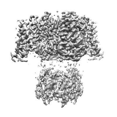 emd_25416.png
 http://ftp.pdbj.org/pub/emdb/structures/EMD-25416


 Z (Sec.)
 Y (Row.)
 X (Col.)


 Sample components
 Processing
 Electron microscopy
 FIELD EMISSION GUN

