National Institutes of Health/National Institute of General Medical Sciences (NIH/NIGMS)
R35 GM127034
United States
Estonian Research Council
PUTJD906
Estonia
National Institutes of Health/National Institute of General Medical Sciences (NIH/NIGMS)
R01 GM121858
United States
National Institutes of Health/National Institute of General Medical Sciences (NIH/NIGMS)
R01 GM131626
United States
National Institutes of Health/National Institute of General Medical Sciences (NIH/NIGMS)
R35 GM139564
United States
National Institutes of Health/National Institute of General Medical Sciences (NIH/NIGMS)
R35 GM139654
United States
National Institutes of Health/National Institute of General Medical Sciences (NIH/NIGMS)
T32 GM086252
United States
National Science Foundation (NSF, United States)
1715321
United States
National Institutes of Health/National Institute of General Medical Sciences (NIH/NIGMS)
U24GM129547
United States
Citation
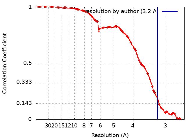
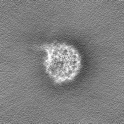
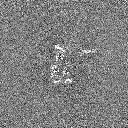
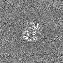
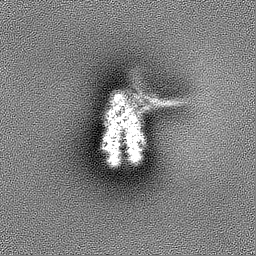
Journal: Mol Cell / Year: 2023 Title: Basic helix-loop-helix pioneer factors interact with the histone octamer to invade nucleosomes and generate nucleosome-depleted regions. Authors: Benjamin T Donovan / Hengye Chen / Priit Eek / Zhiyuan Meng / Caroline Jipa / Song Tan / Lu Bai / Michael G Poirier / Abstract: Nucleosomes drastically limit transcription factor (TF) occupancy, while pioneer transcription factors (PFs) somehow circumvent this nucleosome barrier. In this study, we compare nucleosome binding ...Nucleosomes drastically limit transcription factor (TF) occupancy, while pioneer transcription factors (PFs) somehow circumvent this nucleosome barrier. In this study, we compare nucleosome binding of two conserved S. cerevisiae basic helix-loop-helix (bHLH) TFs, Cbf1 and Pho4. A cryo-EM structure of Cbf1 in complex with the nucleosome reveals that the Cbf1 HLH region can electrostatically interact with exposed histone residues within a partially unwrapped nucleosome. Single-molecule fluorescence studies show that the Cbf1 HLH region facilitates efficient nucleosome invasion by slowing its dissociation rate relative to DNA through interactions with histones, whereas the Pho4 HLH region does not. In vivo studies show that this enhanced binding provided by the Cbf1 HLH region enables nucleosome invasion and ensuing repositioning. These structural, single-molecule, and in vivo studies reveal the mechanistic basis of dissociation rate compensation by PFs and how this translates to facilitating chromatin opening inside cells.
In the structure databanks used in Yorodumi, some data are registered as the other names, "COVID-19 virus" and "2019-nCoV". Here are the details of the virus and the list of structure data.
Jan 31, 2019. EMDB accession codes are about to change! (news from PDBe EMDB page)
EMDB accession codes are about to change! (news from PDBe EMDB page)
The allocation of 4 digits for EMDB accession codes will soon come to an end. Whilst these codes will remain in use, new EMDB accession codes will include an additional digit and will expand incrementally as the available range of codes is exhausted. The current 4-digit format prefixed with “EMD-” (i.e. EMD-XXXX) will advance to a 5-digit format (i.e. EMD-XXXXX), and so on. It is currently estimated that the 4-digit codes will be depleted around Spring 2019, at which point the 5-digit format will come into force.
The EM Navigator/Yorodumi systems omit the EMD- prefix.
Related info.:Q: What is EMD? / ID/Accession-code notation in Yorodumi/EM Navigator
Yorodumi is a browser for structure data from EMDB, PDB, SASBDB, etc.
This page is also the successor to EM Navigator detail page, and also detail information page/front-end page for Omokage search.
The word "yorodu" (or yorozu) is an old Japanese word meaning "ten thousand". "mi" (miru) is to see.
Related info.:EMDB / PDB / SASBDB / Comparison of 3 databanks / Yorodumi Search / Aug 31, 2016. New EM Navigator & Yorodumi / Yorodumi Papers / Jmol/JSmol / Function and homology information / Changes in new EM Navigator and Yorodumi
 Movie
Movie Controller

 Yorodumi
 Open data
 Basic information
 Map data
 Sample
 Keywords
 Function and homology information
 Authors
 United States,
  Estonia, 9 items
  Citation
 Structure visualization
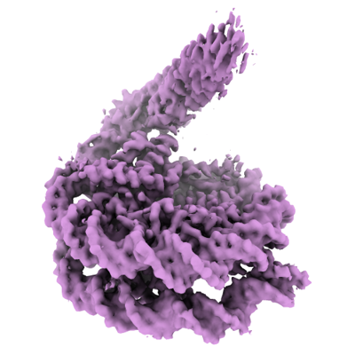
 Downloads & links
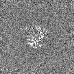
 emd_25406.png
 http://ftp.pdbj.org/pub/emdb/structures/EMD-25406


 Z (Sec.)
 Y (Row.)
 X (Col.)


 Sample components
 Processing
 Electron microscopy
 FIELD EMISSION GUN