 Movie
Movie Controller

[English] 日本語
 Yorodumi
- EMDB-24775: Cryo-EM structure of the SARS-CoV-2 HR1HR2 fusion core complex wi... -
+ Open data

- Basic information
| Entry |  | |||||||||
|---|---|---|---|---|---|---|---|---|---|---|
| Title | Cryo-EM structure of the SARS-CoV-2 HR1HR2 fusion core complex with D936Y mutation | |||||||||
 Map data Map data | ||||||||||
 Sample Sample |
| |||||||||
 Keywords Keywords | spike / HR1HR2 / fusion / D936Y / VIRAL PROTEIN | |||||||||
| Function / homology |  Function and homology information Function and homology informationoxidoreductase activity, acting on metal ions / ferric iron binding / symbiont-mediated disruption of host tissue / Maturation of spike protein / Translation of Structural Proteins / Virion Assembly and Release / host cell surface / host extracellular region / symbiont-mediated-mediated suppression of host tetherin activity / Induction of Cell-Cell Fusion ...oxidoreductase activity, acting on metal ions / ferric iron binding / symbiont-mediated disruption of host tissue / Maturation of spike protein / Translation of Structural Proteins / Virion Assembly and Release / host cell surface / host extracellular region / symbiont-mediated-mediated suppression of host tetherin activity / Induction of Cell-Cell Fusion / structural constituent of virion / positive regulation of viral entry into host cell / membrane fusion / host cell endoplasmic reticulum-Golgi intermediate compartment membrane / Attachment and Entry / entry receptor-mediated virion attachment to host cell / receptor-mediated virion attachment to host cell / host cell surface receptor binding / symbiont-mediated suppression of host innate immune response / endocytosis involved in viral entry into host cell / receptor ligand activity / fusion of virus membrane with host plasma membrane / fusion of virus membrane with host endosome membrane / viral envelope / symbiont entry into host cell / virion attachment to host cell / host cell plasma membrane / SARS-CoV-2 activates/modulates innate and adaptive immune responses / virion membrane / membrane / identical protein binding / plasma membrane Similarity search - Function | |||||||||
| Biological species |   Severe acute respiratory syndrome coronavirus 2 Severe acute respiratory syndrome coronavirus 2 | |||||||||
| Method | single particle reconstruction / cryo EM / Resolution: 2.27 Å | |||||||||
 Authors Authors | Yang K / Brunger AT | |||||||||
| Funding support |  United States, 1 items United States, 1 items
| |||||||||
 Citation Citation | Journal: Proc Natl Acad Sci U S A / Year: 2022 Title: Structural conservation among variants of the SARS-CoV-2 spike postfusion bundle. Authors: Kailu Yang / Chuchu Wang / K Ian White / Richard A Pfuetzner / Luis Esquivies / Axel T Brunger / Abstract: Variants of severe acute respiratory syndrome coronavirus 2 (SARS-CoV-2) challenge currently available COVID-19 vaccines and monoclonal antibody therapies due to structural and dynamic changes of the ...Variants of severe acute respiratory syndrome coronavirus 2 (SARS-CoV-2) challenge currently available COVID-19 vaccines and monoclonal antibody therapies due to structural and dynamic changes of the viral spike glycoprotein (S). The heptad repeat 1 (HR1) and heptad repeat 2 (HR2) domains of S drive virus–host membrane fusion by assembly into a six-helix bundle, resulting in delivery of viral RNA into the host cell. We surveyed mutations of currently reported SARS-CoV-2 variants and selected eight mutations, including Q954H, N969K, and L981F from the Omicron variant, in the postfusion HR1HR2 bundle for functional and structural studies. We designed a molecular scaffold to determine cryogenic electron microscopy (cryo-EM) structures of HR1HR2 at 2.2–3.8 Å resolution by linking the trimeric N termini of four HR1 fragments to four trimeric C termini of the Dps4 dodecamer from Nostoc punctiforme. This molecular scaffold enables efficient sample preparation and structure determination of the HR1HR2 bundle and its mutants by single-particle cryo-EM. Our structure of the wild-type HR1HR2 bundle resolves uncertainties in previously determined structures. The mutant structures reveal side-chain positions of the mutations and their primarily local effects on the interactions between HR1 and HR2. These mutations do not alter the global architecture of the postfusion HR1HR2 bundle, suggesting that the interfaces between HR1 and HR2 are good targets for developing antiviral inhibitors that should be efficacious against all known variants of SARS-CoV-2 to date. We also note that this work paves the way for similar studies in more distantly related viruses. | |||||||||
| History |
|
- Structure visualization
| Supplemental images |
|---|

- Downloads & links
-EMDB archive
| Map data | emd_24775.map.gz | 5.6 MB | EMDB map data format | |
|---|---|---|---|---|
| Header (meta data) | emd-24775-v30.xmlemd-24775.xml | 10.6 KB 10.6 KB | Display Display | EMDB header |
| Images | 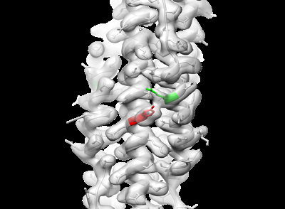 emd_24775.png emd_24775.png | 65.5 KB | ||
| Filedesc metadata | emd-24775.cif.gz | 5.1 KB | ||
| Archive directory |  http://ftp.pdbj.org/pub/emdb/structures/EMD-24775ftp://ftp.pdbj.org/pub/emdb/structures/EMD-24775 http://ftp.pdbj.org/pub/emdb/structures/EMD-24775ftp://ftp.pdbj.org/pub/emdb/structures/EMD-24775 | HTTPS FTP |
-Related structure data
| Related structure data |  7rzrMC  7rzqC  7rzsC  7rztC  7rzuC  7rzvC C: citing same article ( M: atomic model generated by this map |
|---|---|
| Similar structure data |


-Links
| EMDB pages | EMDB (EBI/PDBe) / EMDataResource |
|---|---|
| Related items in Molecule of the Month |


-Map
| File | Download / File: emd_24775.map.gz / Format: CCP4 / Size: 64 MB / Type: IMAGE STORED AS FLOATING POINT NUMBER (4 BYTES) | ||||||||||||||||||||||||||||||||||||
|---|---|---|---|---|---|---|---|---|---|---|---|---|---|---|---|---|---|---|---|---|---|---|---|---|---|---|---|---|---|---|---|---|---|---|---|---|---|
| Projections & slices | Image control
Images are generated by Spider. | ||||||||||||||||||||||||||||||||||||
| Voxel size | X=Y=Z: 0.81625 Å | ||||||||||||||||||||||||||||||||||||
| Density |
| ||||||||||||||||||||||||||||||||||||
| Symmetry | Space group: 1 | ||||||||||||||||||||||||||||||||||||
| Details | EMDB XML:
|
 Z (Sec.)
 Y (Row.)
 X (Col.)


-Supplemental data
- Sample components
-Entire : SARS-CoV-2 HR1HR2 complex with D936Y mutation
| Entire | Name: SARS-CoV-2 HR1HR2 complex with D936Y mutation |
|---|---|
| Components |
|
-Supramolecule #1: SARS-CoV-2 HR1HR2 complex with D936Y mutation
| Supramolecule | Name: SARS-CoV-2 HR1HR2 complex with D936Y mutation / type: complex / ID: 1 / Parent: 0 / Macromolecule list: all |
|---|---|
| Source (natural) | Organism: Severe acute respiratory syndrome coronavirus 2 |
| Molecular weight | Theoretical: 40 KDa |
-Macromolecule #1: SARS-CoV-2 HR1 D936Y linked to a scaffold,Spike protein S2'
| Macromolecule | Name: SARS-CoV-2 HR1 D936Y linked to a scaffold,Spike protein S2' type: protein_or_peptide / ID: 1 / Number of copies: 3 / Enantiomer: LEVO |
|---|---|
| Source (natural) | Organism: Severe acute respiratory syndrome coronavirus 2 |
| Molecular weight | Theoretical: 28.807141 KDa |
| Recombinant expression | Organism:  |
| Sequence | String: MSHHHHHHGS QTLLRNFGNV YDNPVLLDRS VTAPVTEGFN VVLASFQALY LQYQKHHFVV EGSEFYSLHE FFNESYNQVQ DHIHEIGER LDGLGGVPVA TFSKLAELTC FEQESEGVYS SRQMVENDLA AEQAIIGVIR RQAAQAESLG DRGTRYLYEK I LLKTEERA ...String: MSHHHHHHGS QTLLRNFGNV YDNPVLLDRS VTAPVTEGFN VVLASFQALY LQYQKHHFVV EGSEFYSLHE FFNESYNQVQ DHIHEIGER LDGLGGVPVA TFSKLAELTC FEQESEGVYS SRQMVENDLA AEQAIIGVIR RQAAQAESLG DRGTRYLYEK I LLKTEERA YHLSHFLAKD SLTLGFAYEN QKLIANQFNS AIGKIQYSLS STASALGKLQ DVVNQNAQAL NTLVKQLSSN FG AISSVLN DILSRLDKVE UniProtKB: Ferritin, Dps family protein, Spike glycoprotein |
-Macromolecule #2: Spike protein S2'
| Macromolecule | Name: Spike protein S2' / type: protein_or_peptide / ID: 2 / Number of copies: 3 / Enantiomer: LEVO |
|---|---|
| Source (natural) | Organism: Severe acute respiratory syndrome coronavirus 2 |
| Molecular weight | Theoretical: 4.422881 KDa |
| Recombinant expression | Organism: |
| Sequence | String: GPDVDLGDIS GINASVVNIQ KEIDRLNEVA KNLNESLIDL Q UniProtKB: Spike glycoprotein |
-Experimental details
-Structure determination
| Method | cryo EM |
|---|---|
 Processing Processing | single particle reconstruction |
| Aggregation state | particle |
-Sample preparation
| Buffer | pH: 7.4 |
|---|---|
| Vitrification | Cryogen name: ETHANE |
- Electron microscopy
| Microscope | FEI TITAN KRIOS |
|---|---|
| Image recording | Film or detector model: GATAN K3 (6k x 4k) / Average electron dose: 51.0 e/Å2 |
| Electron beam | Acceleration voltage: 300 kV / Electron source:  FIELD EMISSION GUN FIELD EMISSION GUN |
| Electron optics | Illumination mode: FLOOD BEAM / Imaging mode: BRIGHT FIELD |
| Experimental equipment |  Model: Titan Krios / Image courtesy: FEI Company |
-Image processing
| Startup model | Type of model: NONE |
|---|---|
| Final reconstruction | Resolution.type: BY AUTHOR / Resolution: 2.27 Å / Resolution method: FSC 0.143 CUT-OFF / Number images used: 585122 |
| Initial angle assignment | Type: MAXIMUM LIKELIHOOD |
| Final angle assignment | Type: MAXIMUM LIKELIHOOD |
