 Movie
Movie Controller

[English] 日本語
 Yorodumi
- EMDB-24603: Tomogram of SARS-CoV-2 spike-bearing virus-like particles (VLPs) ... -
+ Open data

- Basic information
| Entry | 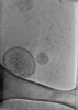 | |||||||||
|---|---|---|---|---|---|---|---|---|---|---|
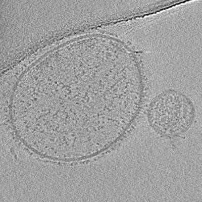
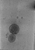
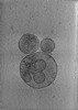
| Title | Tomogram of SARS-CoV-2 spike-bearing virus-like particles (VLPs) interacting with hACE2-bearing extracellular vesicles (tEVs), showing various intermediate states of the SARS-CoV-2 spike protein (Fig. 3D,E of the manuscript Marcink et al., 2021). | |||||||||
 Map data Map data | Tomogram of SARS-CoV-2 spike-bearing virus-like particles (VLPs) interacting with hACE2-bearing extracellular vesicles (tEVs), showing various intermediate states of the SARS-CoV-2 spike protein (Fig. 3D,E of the manuscript Marcink et al., 2021). | |||||||||
 Sample Sample |
| |||||||||
| Biological species |   Severe acute respiratory syndrome coronavirus 2 / Severe acute respiratory syndrome coronavirus 2 /  Homo sapiens HEK293-T (human) Homo sapiens HEK293-T (human) | |||||||||
| Method | electron tomography / cryo EM | |||||||||
 Authors Authors | Marcink TC / Porotto M / des Georges A / Moscona A | |||||||||
| Funding support |  United States, 2 items United States, 2 items
| |||||||||
 Citation Citation | Journal: Sci Adv / Year: 2022 Title: Intermediates in SARS-CoV-2 spike-mediated cell entry. Authors: Tara C Marcink / Thomas Kicmal / Emily Armbruster / Zhening Zhang / Gillian Zipursky / Kate L Golub / Mohab Idris / Jonathan Khao / Jennifer Drew-Bear / Gael McGill / Tom Gallagher / Matteo ...Authors: Tara C Marcink / Thomas Kicmal / Emily Armbruster / Zhening Zhang / Gillian Zipursky / Kate L Golub / Mohab Idris / Jonathan Khao / Jennifer Drew-Bear / Gael McGill / Tom Gallagher / Matteo Porotto / Amédée des Georges / Anne Moscona /  Abstract: SARS-CoV-2 cell entry is completed after viral spike (S) protein-mediated membrane fusion between viral and host cell membranes. Stable prefusion and postfusion S structures have been resolved by ...SARS-CoV-2 cell entry is completed after viral spike (S) protein-mediated membrane fusion between viral and host cell membranes. Stable prefusion and postfusion S structures have been resolved by cryo-electron microscopy and cryo-electron tomography, but the refolding intermediates on the fusion pathway are transient and have not been examined. We used an antiviral lipopeptide entry inhibitor to arrest S protein refolding and thereby capture intermediates as S proteins interact with hACE2 and fusion-activating proteases on cell-derived target membranes. Cryo-electron tomography imaged both extended and partially folded intermediate states of S2, as well as a novel late-stage conformation on the pathway to membrane fusion. The intermediates now identified in this dynamic S protein-directed fusion provide mechanistic insights that may guide the design of CoV entry inhibitors. | |||||||||
| History |
|
- Structure visualization
| Supplemental images |
|---|
- Downloads & links
-EMDB archive
| Map data | emd_24603.map.gz | 1.2 GB |  EMDB map data format EMDB map data format | |
|---|---|---|---|---|
| Header (meta data) | emd-24603-v30.xmlemd-24603.xml | 12 KB 12 KB | Display Display | EMDB header |
| Images |  emd_24603.png emd_24603.png | 180.8 KB | ||
| Archive directory |  http://ftp.pdbj.org/pub/emdb/structures/EMD-24603ftp://ftp.pdbj.org/pub/emdb/structures/EMD-24603 http://ftp.pdbj.org/pub/emdb/structures/EMD-24603ftp://ftp.pdbj.org/pub/emdb/structures/EMD-24603 | HTTPS FTP |
-Related structure data

-Links
| EMDB pages | EMDB (EBI/PDBe) / EMDataResource |
|---|
-Map
| File | Download / File: emd_24603.map.gz / Format: CCP4 / Size: 1.4 GB / Type: IMAGE STORED AS FLOATING POINT NUMBER (4 BYTES) | ||||||||||||||||||||||||||||||||
|---|---|---|---|---|---|---|---|---|---|---|---|---|---|---|---|---|---|---|---|---|---|---|---|---|---|---|---|---|---|---|---|---|---|
| Annotation | Tomogram of SARS-CoV-2 spike-bearing virus-like particles (VLPs) interacting with hACE2-bearing extracellular vesicles (tEVs), showing various intermediate states of the SARS-CoV-2 spike protein (Fig. 3D,E of the manuscript Marcink et al., 2021). | ||||||||||||||||||||||||||||||||
| Projections & slices | Image control
Images are generated by Spider. generated in cubic-lattice coordinate | ||||||||||||||||||||||||||||||||
| Voxel size | X=Y=Z: 6.484 Å | ||||||||||||||||||||||||||||||||
| Density |
| ||||||||||||||||||||||||||||||||
| Symmetry | Space group: 1 | ||||||||||||||||||||||||||||||||
| Details | EMDB XML:
|
 Z (Sec.)
 Y (Row.)
 X (Col.)


-Supplemental data
- Sample components
-Entire : Virus-like particles and extracellular vesicles
| Entire | Name: Virus-like particles and extracellular vesicles |
|---|---|
| Components |
|
-Supramolecule #1: Virus-like particles and extracellular vesicles
| Supramolecule | Name: Virus-like particles and extracellular vesicles / type: complex / ID: 1 / Parent: 0 |
|---|
-Supramolecule #2: hACE2-bearing target extracellular vesicles(tEVs)
| Supramolecule | Name: hACE2-bearing target extracellular vesicles(tEVs) / type: complex / ID: 2 / Parent: 1 |
|---|---|
| Source (natural) | Organism: Severe acute respiratory syndrome coronavirus 2 |
-Supramolecule #3: SARS-CoV-2 spike-bearing virus-like particles (VLPs)
| Supramolecule | Name: SARS-CoV-2 spike-bearing virus-like particles (VLPs) / type: complex / ID: 3 / Parent: 1 |
|---|---|
| Source (natural) | Organism: Homo sapiens HEK293-T (human) |
-Experimental details
-Structure determination
| Method | cryo EM |
|---|---|
 Processing Processing | electron tomography |
| Aggregation state | particle |
-Sample preparation
| Buffer | pH: 7.4 |
|---|---|
| Grid | Model: PELCO Ultrathin Carbon with Lacey Carbon / Material: GOLD / Mesh: 300 / Support film - Material: CARBON / Support film - topology: LACEY / Support film - Film thickness: 300.0 nm |
| Vitrification | Cryogen name: ETHANE / Chamber humidity: 100 % / Chamber temperature: 277.15 K / Instrument: FEI VITROBOT MARK IV |
| Sectioning | Other: NO SECTIONING |
| Fiducial marker | Manufacturer: Sigma Aldrich / Diameter: 5 nm |
- Electron microscopy
| Microscope | FEI TITAN KRIOS |
|---|---|
| Image recording | Film or detector model: GATAN K3 BIOQUANTUM (6k x 4k) / Average electron dose: 2.8 e/Å2 |
| Electron beam | Acceleration voltage: 300 kV / Electron source:  FIELD EMISSION GUN FIELD EMISSION GUN |
| Electron optics | Calibrated defocus max: 8.0 µm / Calibrated defocus min: 5.0 µm / Calibrated magnification: 53000 / Illumination mode: FLOOD BEAM / Imaging mode: BRIGHT FIELD |
| Sample stage | Specimen holder model: FEI TITAN KRIOS AUTOGRID HOLDER |
| Experimental equipment |  Model: Titan Krios / Image courtesy: FEI Company |
-Image processing
| Final reconstruction | Algorithm: BACK PROJECTION / Software - Name: eTomo (ver. 4.10.52) / Number images used: 37 |
|---|---|
| CTF correction | Software: (Name: Warp (ver. 1.0.9), eTomo (ver. 4.10.52)) |
