 ムービー
ムービー コントローラー


+ データを開く

- 基本情報
基本情報
| 登録情報 | データベース: EMDB / ID: EMD-24433 | |||||||||
|---|---|---|---|---|---|---|---|---|---|---|
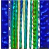
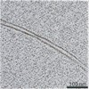
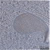
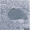
| タイトル | Yeast Nucleus Tomogram | |||||||||
 マップデータ マップデータ | ||||||||||
 試料 試料 |
| |||||||||
| 生物種 |  | |||||||||
| 手法 | 電子線トモグラフィー法 / クライオ電子顕微鏡法 | |||||||||
 データ登録者 データ登録者 | Croxford M / Villa E | |||||||||
| 資金援助 |  米国, 1件 米国, 1件
| |||||||||
 引用 引用 | ジャーナル: Proc Natl Acad Sci U S A / 年: 2021 タイトル: Entropy-regularized deconvolution of cellular cryotransmission electron tomograms. 著者: Matthew Croxford / Michael Elbaum / Muthuvel Arigovindan / Zvi Kam / David Agard / Elizabeth Villa / John Sedat /   要旨: Cryo-electron tomography (cryo-ET) allows for the high-resolution visualization of biological macromolecules. However, the technique is limited by a low signal-to-noise ratio (SNR) and variance in ...Cryo-electron tomography (cryo-ET) allows for the high-resolution visualization of biological macromolecules. However, the technique is limited by a low signal-to-noise ratio (SNR) and variance in contrast at different frequencies, as well as reduced Z resolution. Here, we applied entropy-regularized deconvolution (ER-DC) to cryo-ET data generated from transmission electron microscopy (TEM) and reconstructed using weighted back projection (WBP). We applied deconvolution to several in situ cryo-ET datasets and assessed the results by Fourier analysis and subtomogram analysis (STA). | |||||||||
| 履歴 |
|
- 構造の表示
構造の表示
| ムービー |
 ムービービューア ムービービューア |
|---|---|
| 添付画像 |

- ダウンロードとリンク
-EMDBアーカイブ
| マップデータ | emd_24433.map.gz | 1.1 GB | EMDBマップデータ形式 | |
|---|---|---|---|---|
| ヘッダ (付随情報) | emd-24433-v30.xmlemd-24433.xml | 8 KB 8 KB | 表示 表示 | EMDBヘッダ |
| 画像 | 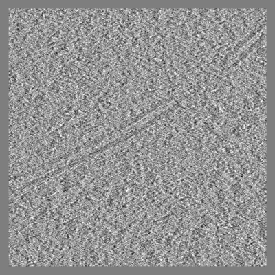 emd_24433.png emd_24433.png | 215.7 KB | ||
| アーカイブディレクトリ |  http://ftp.pdbj.org/pub/emdb/structures/EMD-24433ftp://ftp.pdbj.org/pub/emdb/structures/EMD-24433 http://ftp.pdbj.org/pub/emdb/structures/EMD-24433ftp://ftp.pdbj.org/pub/emdb/structures/EMD-24433 | HTTPS FTP |
-検証レポート
| 文書・要旨 | emd_24433_validation.pdf.gz | 320.9 KB | 表示 | EMDB検証レポート |
|---|---|---|---|---|
| 文書・詳細版 | emd_24433_full_validation.pdf.gz | 320.4 KB | 表示 | |
| XML形式データ | emd_24433_validation.xml.gz | 4.3 KB | 表示 | |
| CIF形式データ | emd_24433_validation.cif.gz | 4.7 KB | 表示 | |
| アーカイブディレクトリ | https://ftp.pdbj.org/pub/emdb/validation_reports/EMD-24433ftp://ftp.pdbj.org/pub/emdb/validation_reports/EMD-24433 | HTTPS FTP |
-関連構造データ
| 関連構造データ | C: 同じ文献を引用 ( |
|---|---|
| 電子顕微鏡画像生データ | EMPIAR-10762 (タイトル: Entropy Regularized Deconvolution of Cellular Cryo-Transmission Electron Tomograms Data size: 5.2 Data #1: Aligned tilt series, and corresponding deconvolution of a yeast nucleus [tilt series]) |
-リンク
| EMDBのページ | EMDB (EBI/PDBe) / EMDataResource |
|---|
-マップ
| ファイル | ダウンロード / ファイル: emd_24433.map.gz / 形式: CCP4 / 大きさ: 1.2 GB / タイプ: IMAGE STORED AS FLOATING POINT NUMBER (4 BYTES) | ||||||||||||||||||||||||||||||||||||||||||||||||||||||||||||
|---|---|---|---|---|---|---|---|---|---|---|---|---|---|---|---|---|---|---|---|---|---|---|---|---|---|---|---|---|---|---|---|---|---|---|---|---|---|---|---|---|---|---|---|---|---|---|---|---|---|---|---|---|---|---|---|---|---|---|---|---|---|
| ボクセルのサイズ | X=Y=Z: 10.62 Å | ||||||||||||||||||||||||||||||||||||||||||||||||||||||||||||
| 密度 |
| ||||||||||||||||||||||||||||||||||||||||||||||||||||||||||||
| 対称性 | 空間群: 1 | ||||||||||||||||||||||||||||||||||||||||||||||||||||||||||||
| 詳細 | EMDB XML:
CCP4マップ ヘッダ情報:
| ||||||||||||||||||||||||||||||||||||||||||||||||||||||||||||
-添付データ
- 試料の構成要素
-全体 : Yeast Nuclear Periphery and Adjacent Cytosol
| 全体 | 名称: Yeast Nuclear Periphery and Adjacent Cytosol |
|---|---|
| 要素 |
|
-超分子 #1: Yeast Nuclear Periphery and Adjacent Cytosol
| 超分子 | 名称: Yeast Nuclear Periphery and Adjacent Cytosol / タイプ: cell / ID: 1 / 親要素: 0 |
|---|---|
| 由来(天然) | 生物種: |
-実験情報
-構造解析
| 手法 | クライオ電子顕微鏡法 |
|---|---|
 解析 解析 | 電子線トモグラフィー法 |
| 試料の集合状態 | cell |
-試料調製
| 緩衝液 | pH: 7.4 |
|---|---|
| グリッド | モデル: Quantifoil / 材質: COPPER |
| 凍結 | 凍結剤: ETHANE-PROPANE |
| 切片作成 | 集束イオンビーム - 装置: OTHER / 集束イオンビーム - イオン: OTHER / 集束イオンビーム - 電圧: 30 kV / 集束イオンビーム - 電流: 0.03 nA / 集束イオンビーム - 時間: 1000 sec. / 集束イオンビーム - 温度: 83 K / 集束イオンビーム - Initial thickness: 4000 nm / 集束イオンビーム - 最終 厚さ: 350 nm 集束イオンビーム - 詳細: The value given for _emd_sectioning_focused_ion_beam.instrument is FEI Scios FIB. This is not in a list of allowed values {'DB235', 'OTHER'} so OTHER is written into the XML file. |
- 電子顕微鏡法
| 顕微鏡 | FEI POLARA 300 |
|---|---|
| 撮影 | フィルム・検出器のモデル: GATAN K2 SUMMIT (4k x 4k) 平均電子線量: 100.0 e/Å2 |
| 電子線 | 加速電圧: 300 kV / 電子線源:  FIELD EMISSION GUN FIELD EMISSION GUN |
| 電子光学系 | 照射モード: FLOOD BEAM / 撮影モード: BRIGHT FIELD |
| 試料ステージ | ホルダー冷却材: NITROGEN |
| 実験機器 |  モデル: Tecnai Polara / 画像提供: FEI Company |
-画像解析
| 最終 再構成 | アルゴリズム: BACK PROJECTION / 使用した粒子像数: 56 |
|---|
