- EMDB-22334: Human parainfluenza virus fusion complex glycoproteins imaged in ... -
+
データを開く
IDまたはキーワード:
読み込み中...
-
基本情報
登録情報
データベース: EMDB / ID: EMD-22334
タイトル
Human parainfluenza virus fusion complex glycoproteins imaged in action on authentic viral surfaces: Subtomogram average of hemagglutinin-neuraminidase (HN) and fusion (F) protein complex prior to receptor engagement
マップデータ
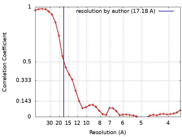
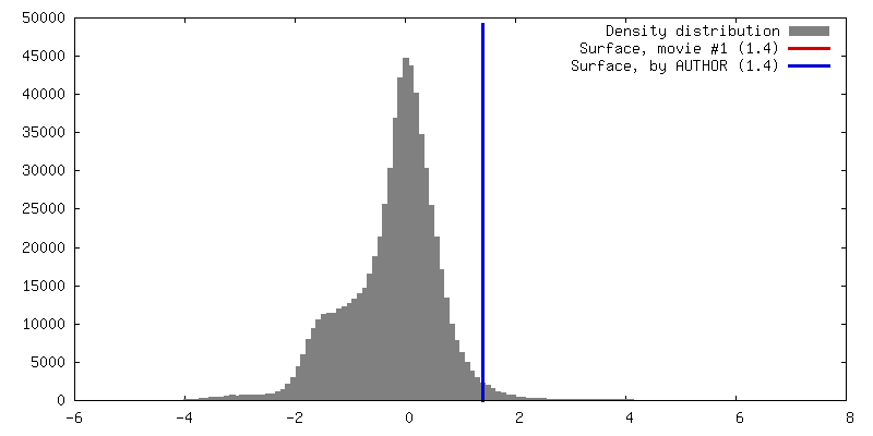
Cryo-electron tomography and subtomogram averaging of the HN and F complex on the surface of HPIV3
National Institutes of Health/National Institute Of Allergy and Infectious Diseases (NIH/NIAID)
RO1AI031971
米国
National Institutes of Health/National Institute Of Allergy and Infectious Diseases (NIH/NIAID)
RO1AI121349
米国
National Institutes of Health/National Institute Of Allergy and Infectious Diseases (NIH/NIAID)
RO1AI114736
米国
National Institutes of Health/National Institute of General Medical Sciences (NIH/NIGMS)
R35GM133598
米国
引用
ジャーナル: mBio / 年: 2020 タイトル: Hijacking the Fusion Complex of Human Parainfluenza Virus as an Antiviral Strategy. 著者: T C Marcink / E Yariv / K Rybkina / V Más / F T Bovier / A des Georges / A L Greninger / C A Alabi / M Porotto / N Ben-Tal / A Moscona / 要旨: The receptor binding protein of parainfluenza virus, hemagglutinin-neuraminidase (HN), is responsible for actively triggering the viral fusion protein (F) to undergo a conformational change leading ...The receptor binding protein of parainfluenza virus, hemagglutinin-neuraminidase (HN), is responsible for actively triggering the viral fusion protein (F) to undergo a conformational change leading to insertion into the target cell and fusion of the virus with the target cell membrane. For proper viral entry to occur, this process must occur when HN is engaged with host cell receptors at the cell surface. It is possible to interfere with this process through premature activation of the F protein, distant from the target cell receptor. Conformational changes in the F protein and adoption of the postfusion form of the protein prior to receptor engagement of HN at the host cell membrane inactivate the virus. We previously identified small molecules that interact with HN and induce it to activate F in an untimely fashion, validating a new antiviral strategy. To obtain highly active pretriggering candidate molecules we carried out a virtual modeling screen for molecules that interact with sialic acid binding site II on HN, which we propose to be the site responsible for activating F. To directly assess the mechanism of action of one such highly effective new premature activating compound, PAC-3066, we use cryo-electron tomography on authentic intact viral particles for the first time to examine the effects of PAC-3066 treatment on the conformation of the viral F protein. We present the first direct observation of the conformational rearrangement induced in the viral F protein. Paramyxoviruses, including human parainfluenza virus type 3, are internalized into host cells by fusion between viral and target cell membranes. The receptor binding protein, hemagglutinin-neuraminidase (HN), upon binding to its cell receptor, triggers conformational changes in the fusion protein (F). This action of HN activates F to reach its fusion-competent state. Using small molecules that interact with HN, we can induce the premature activation of F and inactivate the virus. To obtain highly active pretriggering compounds, we carried out a virtual modeling screen for molecules that interact with a sialic acid binding site on HN that we propose to be the site involved in activating F. We use cryo-electron tomography of authentic intact viral particles for the first time to directly assess the mechanism of action of this treatment on the conformation of the viral F protein and present the first direct observation of the induced conformational rearrangement in the viral F protein.
EMPIAR-10476 (タイトル: Human Parainfluenza Virus Fusion Complex Glycoproteins Imaged In Action On Authentic Viral Surfaces Data size: 11.6 Data #1: Human parainfluenza virus fusion complex glycoproteins imaged in action on authentic viral surfaces: Tomograms of HPIV3 prior to receptor engagement [tilt series])
 ムービー
ムービー コントローラー


 データを開く
 基本情報
基本情報 マップデータ
 試料
試料 Human respirovirus 3 (ウイルス)
 データ登録者
 米国, 4件
  引用
引用


 構造の表示
構造の表示 ムービービューア
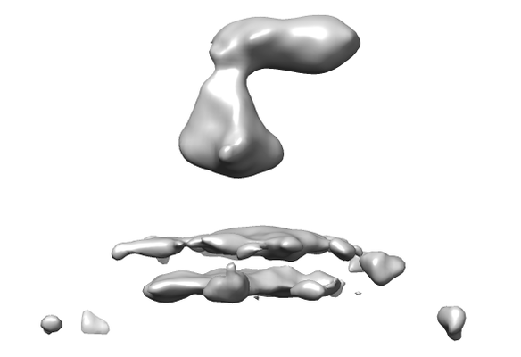
 ダウンロードとリンク
 emd_22334.png
 http://ftp.pdbj.org/pub/emdb/structures/EMD-22334
 Z (Sec.)
 Y (Row.)
 X (Col.)


 試料の構成要素
 Homo sapiens (ヒト)
 Chlorocebus sabaeus (オナガザル)
 解析
解析 電子顕微鏡法
 FIELD EMISSION GUN