 Movie
Movie Controller

[English] 日本語
 Yorodumi
- EMDB-18306: Towards the Visual Proteomics of C. reinhardtii using High-throug... -
+ Open data

- Basic information
| Entry | 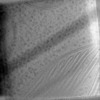 | |||||||||
|---|---|---|---|---|---|---|---|---|---|---|
| Title | Towards the Visual Proteomics of C. reinhardtii using High-throughput Collaborative in situ Cryo-ET : Representative Tomogram | |||||||||
 Map data Map data | ||||||||||
 Sample Sample |
| |||||||||
 Keywords Keywords | Chlamydomonas reinhardtii cryo-Plasma-FIB / PHOTOSYNTHESIS | |||||||||
| Biological species |   Chlamydomonas reinhardtii (plant) Chlamydomonas reinhardtii (plant) | |||||||||
| Method | electron tomography / cryo EM | |||||||||
 Authors Authors | Khavnekar S / Kelley R / Zhang X / Kotecha A | |||||||||
| Funding support |  Germany, 2 items Germany, 2 items
| |||||||||
 Citation Citation | Journal: Microsc Microanal / Year: 2023 Title: Towards the Visual Proteomics of C. reinhardtii using High-throughput Collaborative in situ Cryo-ET. Authors: Sagar Khavnekar / Ron Kelley / Florent Waltz / Wojciech Wietrzynski / Xianjun Zhang / Martin Obr / Grigory Tagiltsev / Florian Beck / William Wan / John Briggs / Ben Engel / Juergen Plitzko / Abhay Kotecha /    | |||||||||
| History |
|
- Structure visualization
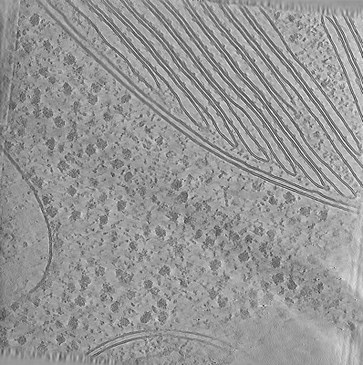
| Supplemental images |
|---|
- Downloads & links
-EMDB archive
| Map data | emd_18306.map.gz | 1.8 GB |  EMDB map data format EMDB map data format | |
|---|---|---|---|---|
| Header (meta data) | emd-18306-v30.xmlemd-18306.xml | 8.2 KB 8.2 KB | Display Display | EMDB header |
| Images |  emd_18306.png emd_18306.png | 224.2 KB | ||
| Filedesc metadata | emd-18306.cif.gz | 3.8 KB | ||
| Archive directory |  http://ftp.pdbj.org/pub/emdb/structures/EMD-18306ftp://ftp.pdbj.org/pub/emdb/structures/EMD-18306 http://ftp.pdbj.org/pub/emdb/structures/EMD-18306ftp://ftp.pdbj.org/pub/emdb/structures/EMD-18306 | HTTPS FTP |
-Links
| EMDB pages | EMDB (EBI/PDBe) / EMDataResource |
|---|
-Map
| File | Download / File: emd_18306.map.gz / Format: CCP4 / Size: 2 GB / Type: IMAGE STORED AS FLOATING POINT NUMBER (4 BYTES) | ||||||||||||||||||||||||||||||||
|---|---|---|---|---|---|---|---|---|---|---|---|---|---|---|---|---|---|---|---|---|---|---|---|---|---|---|---|---|---|---|---|---|---|
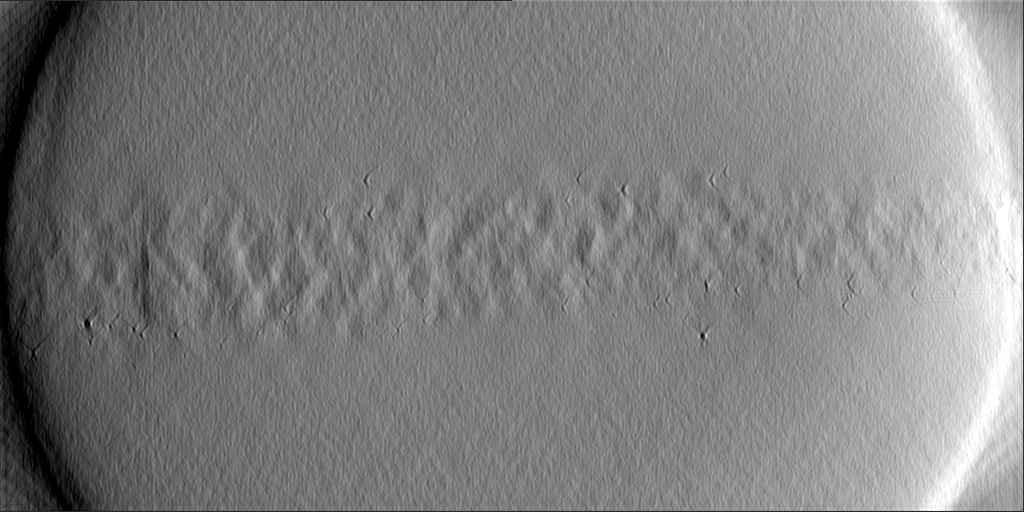
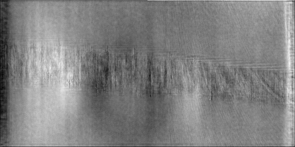
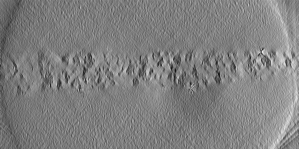
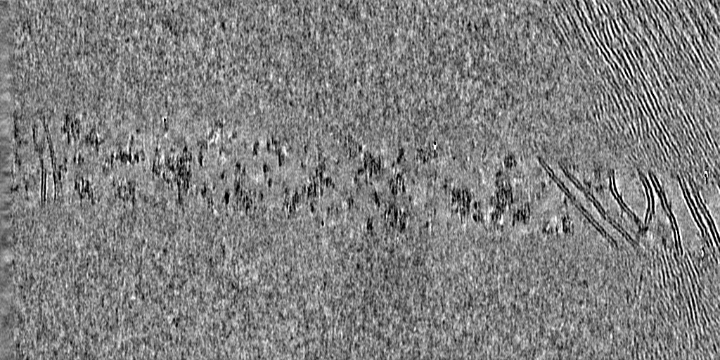
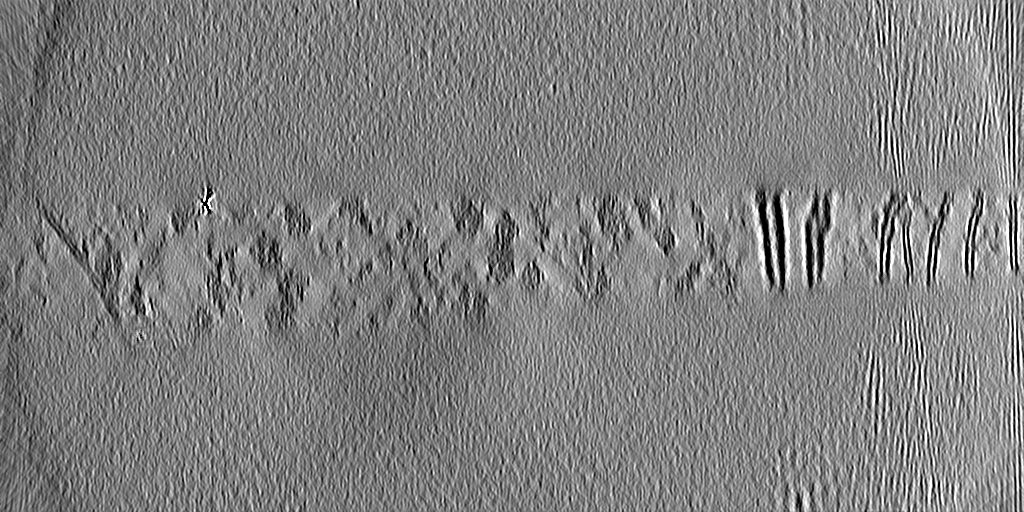
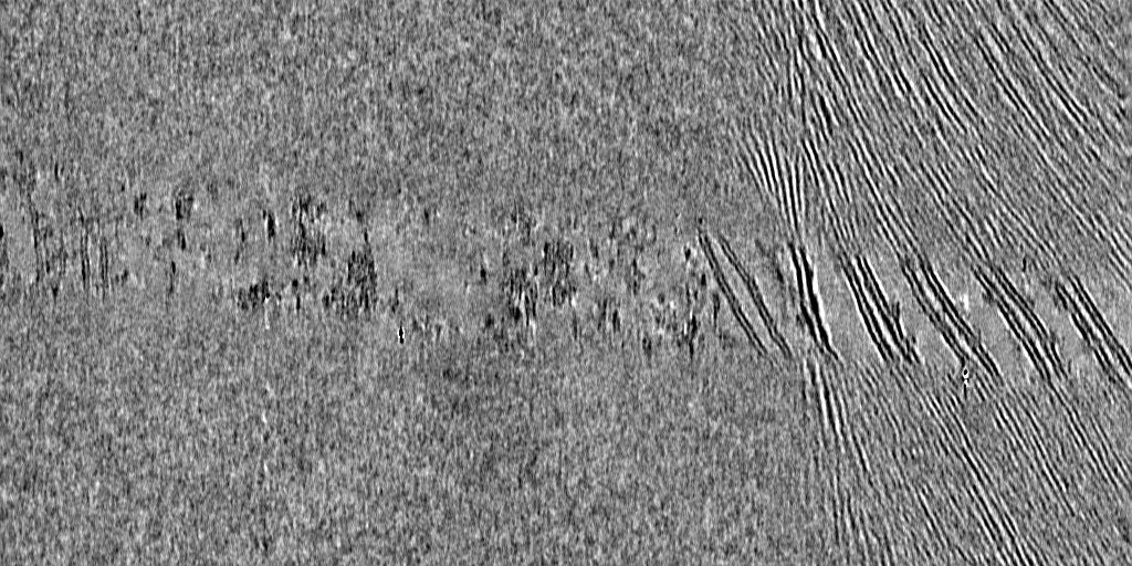
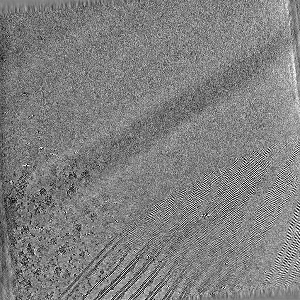
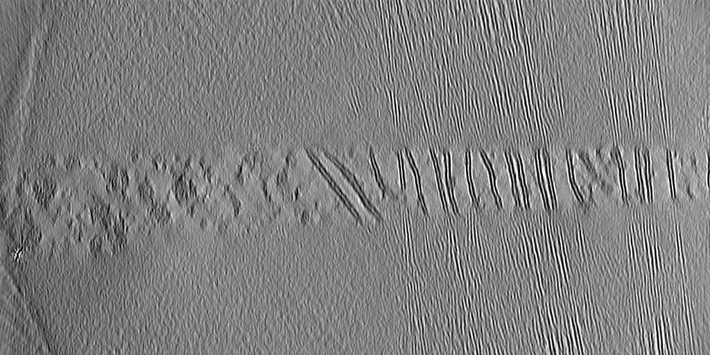
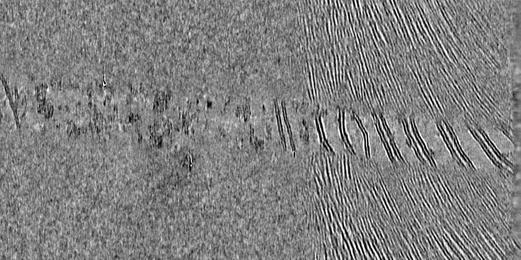
| Projections & slices | Image control
Images are generated by Spider. generated in cubic-lattice coordinate | ||||||||||||||||||||||||||||||||
| Voxel size | X=Y=Z: 7.84 Å | ||||||||||||||||||||||||||||||||
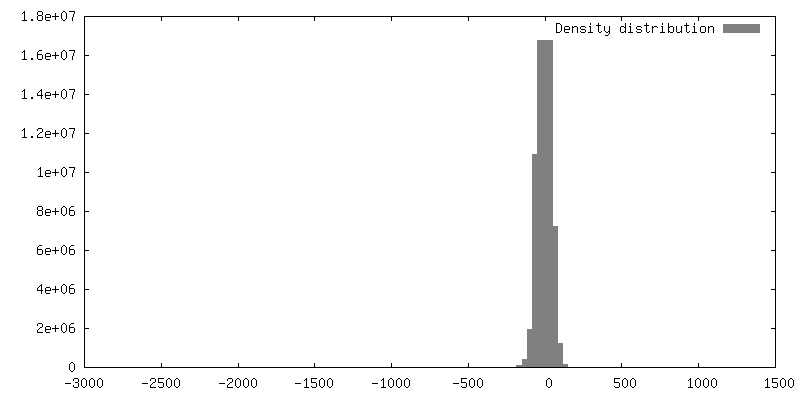
| Density |
| ||||||||||||||||||||||||||||||||
| Symmetry | Space group: 1 | ||||||||||||||||||||||||||||||||
| Details | EMDB XML:
|
 Z (Sec.)
 Y (Row.)
 X (Col.)


-Supplemental data
- Sample components
-Entire : Chlamydomonas reinhardtii
| Entire | Name: Chlamydomonas reinhardtii (plant) |
|---|---|
| Components |
|
-Supramolecule #1: Chlamydomonas reinhardtii
| Supramolecule | Name: Chlamydomonas reinhardtii / type: cell / ID: 1 / Parent: 0 |
|---|---|
| Source (natural) | Organism: Chlamydomonas reinhardtii (plant) / Strain: CC-3994 mat3-4 mt+ |
-Experimental details
-Structure determination
| Method | cryo EM |
|---|---|
 Processing Processing | electron tomography |
| Aggregation state | cell |
-Sample preparation
| Buffer | pH: 7 |
|---|---|
| Vitrification | Cryogen name: ETHANE |
| Sectioning | Focused ion beam - Instrument: OTHER / Focused ion beam - Ion: OTHER / Focused ion beam - Voltage: 12 / Focused ion beam - Current: 0.05 / Focused ion beam - Duration: 120 / Focused ion beam - Temperature: 90 K / Focused ion beam - Initial thickness: 500 / Focused ion beam - Final thickness: 90 Focused ion beam - Details: The value given for _em_focused_ion_beam.instrument is FEI Arctis. This is not in a list of allowed values {'OTHER', 'DB235'} so OTHER is written into the XML file. |
- Electron microscopy
| Microscope | FEI TITAN KRIOS |
|---|---|
| Image recording | Film or detector model: FEI FALCON IV (4k x 4k) / Average electron dose: 3.5 e/Å2 |
| Electron beam | Acceleration voltage: 300 kV / Electron source:  FIELD EMISSION GUN FIELD EMISSION GUN |
| Electron optics | Illumination mode: FLOOD BEAM / Imaging mode: BRIGHT FIELD / Nominal defocus max: 3.5 µm / Nominal defocus min: 1.0 µm |
| Experimental equipment |  Model: Titan Krios / Image courtesy: FEI Company |
-Image processing
| Final reconstruction | Algorithm: BACK PROJECTION / Number images used: 1 |
|---|
