 Movie
Movie Controller

[English] 日本語
 Yorodumi
- EMDB-16537: Optimizing Cryo-FIB Lamellas for sub-5 Angstrom in situ Structura... -
+ Open data

- Basic information
| Entry |  | |||||||||
|---|---|---|---|---|---|---|---|---|---|---|
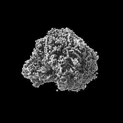
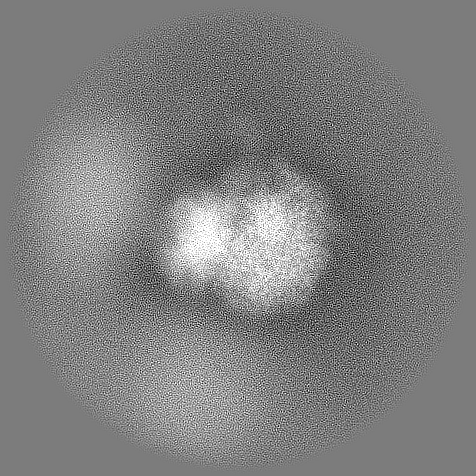
| Title | Optimizing Cryo-FIB Lamellas for sub-5 Angstrom in situ Structural Biology : Subtomogram average of the Large Subunit of S.Cerevisiae 80S Ribosome | |||||||||
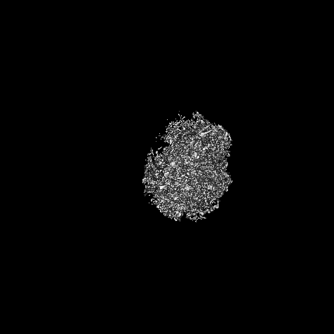
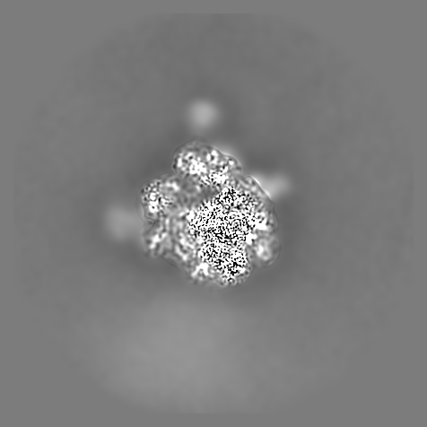
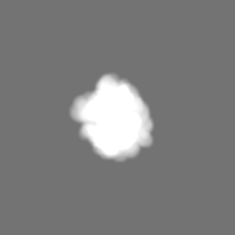
 Map data Map data | Local resolution filtered map after M refinement Workflow: TOMOMAN-STOPGAP-Warp-Relion-M | |||||||||
 Sample Sample |
| |||||||||
 Keywords Keywords | S.Cerevisiae / 80S / Ribosome / FIB / insitu | |||||||||
| Biological species |  | |||||||||
| Method | subtomogram averaging / cryo EM / Resolution: 4.6 Å | |||||||||
 Authors Authors | Khavnekar S / Vrbovska V / Zaoralova M / KElley R / Beck F / Klumpe S / Kotech A / Plitzko JM / Erdmann PS | |||||||||
| Funding support |  Germany, 1 items Germany, 1 items
| |||||||||
 Citation Citation | Journal: To Be Published Title: Optimizing Cryo-FIB Lamellas for sub-5 Angstrom in situ Structural Biology : Large Subunit of S.Cerevisiae 80S Ribosome Authors: Khavnekar S / Vrbovska V / Zaoralova M / KElley R / Beck F / Klumpe S / Kotech A / Plitzko JM / Erdmann PS | |||||||||
| History |
|
- Structure visualization
| Supplemental images |
|---|
- Downloads & links
-EMDB archive
| Map data | emd_16537.map.gz | 222.3 MB |  EMDB map data format EMDB map data format | |
|---|---|---|---|---|
| Header (meta data) | emd-16537-v30.xmlemd-16537.xml | 12.1 KB 12.1 KB | Display Display | EMDB header |
| Images |  emd_16537.png emd_16537.png | 65.3 KB | ||
| Masks | emd_16537_msk_1.map | 411.4 MB | Mask map | |
| Filedesc metadata | emd-16537.cif.gz | 4 KB | ||
| Others | emd_16537_half_map_1.map.gzemd_16537_half_map_2.map.gz | 212.1 MB 212.1 MB | ||
| Archive directory |  http://ftp.pdbj.org/pub/emdb/structures/EMD-16537ftp://ftp.pdbj.org/pub/emdb/structures/EMD-16537 http://ftp.pdbj.org/pub/emdb/structures/EMD-16537ftp://ftp.pdbj.org/pub/emdb/structures/EMD-16537 | HTTPS FTP |
-Links
| EMDB pages | EMDB (EBI/PDBe) / EMDataResource |
|---|
-Map
| File | Download / File: emd_16537.map.gz / Format: CCP4 / Size: 411.4 MB / Type: IMAGE STORED AS FLOATING POINT NUMBER (4 BYTES) | ||||||||||||||||||||||||||||||||||||
|---|---|---|---|---|---|---|---|---|---|---|---|---|---|---|---|---|---|---|---|---|---|---|---|---|---|---|---|---|---|---|---|---|---|---|---|---|---|
| Annotation | Local resolution filtered map after M refinement Workflow: TOMOMAN-STOPGAP-Warp-Relion-M | ||||||||||||||||||||||||||||||||||||
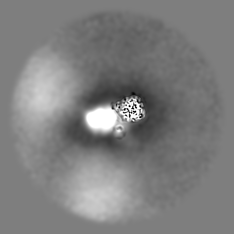
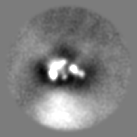
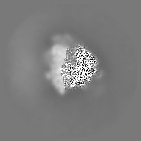
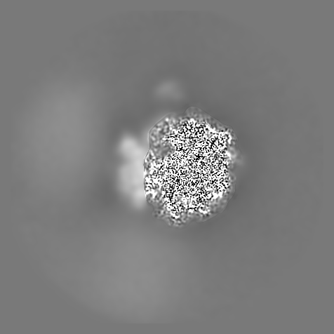
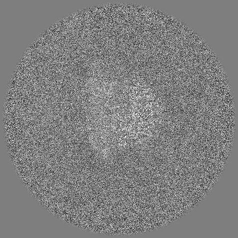
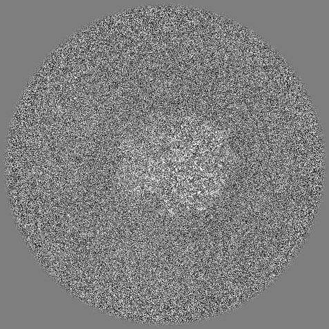
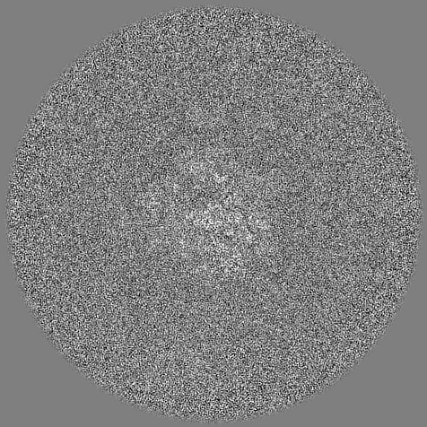
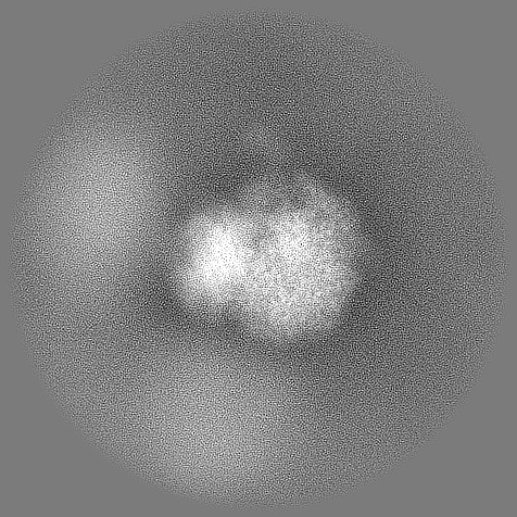
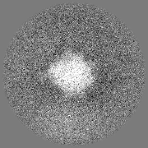
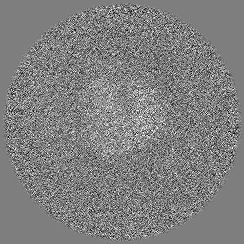
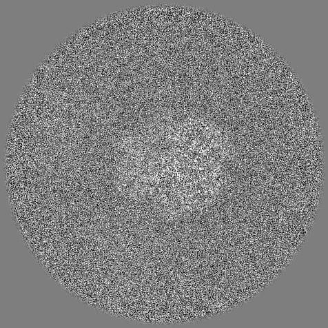
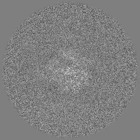
| Projections & slices | Image control
Images are generated by Spider. | ||||||||||||||||||||||||||||||||||||
| Voxel size | X=Y=Z: 1.6 Å | ||||||||||||||||||||||||||||||||||||
| Density |
| ||||||||||||||||||||||||||||||||||||
| Symmetry | Space group: 1 | ||||||||||||||||||||||||||||||||||||
| Details | EMDB XML:
|
 Z (Sec.)
 Y (Row.)
 X (Col.)


-Supplemental data
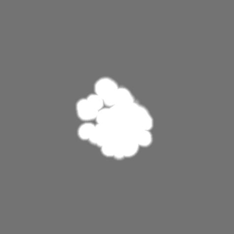
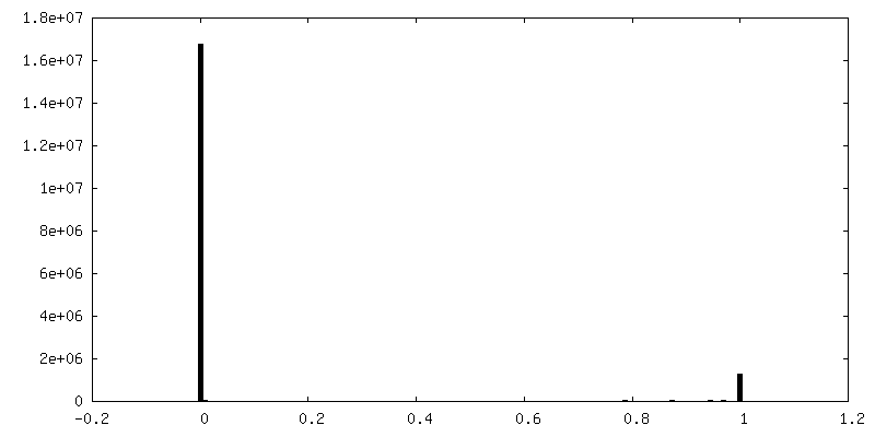
-Mask #1
| File | emd_16537_msk_1.map | ||||||||||||
|---|---|---|---|---|---|---|---|---|---|---|---|---|---|
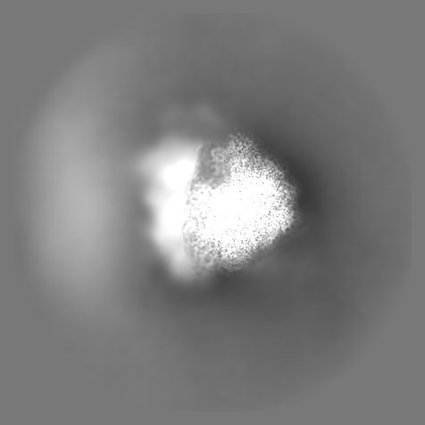
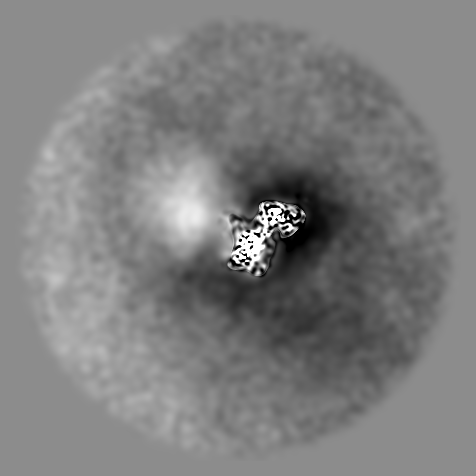
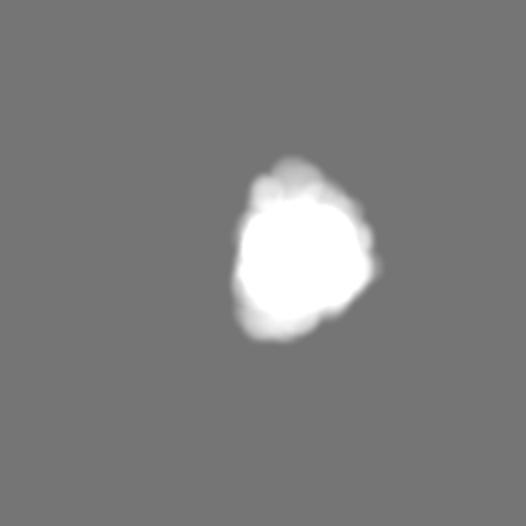
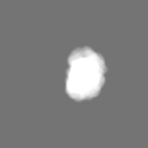
| Projections & Slices |
| ||||||||||||
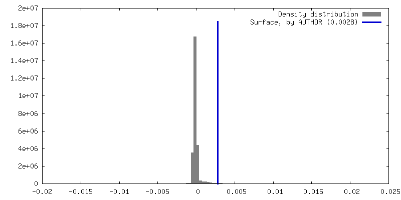
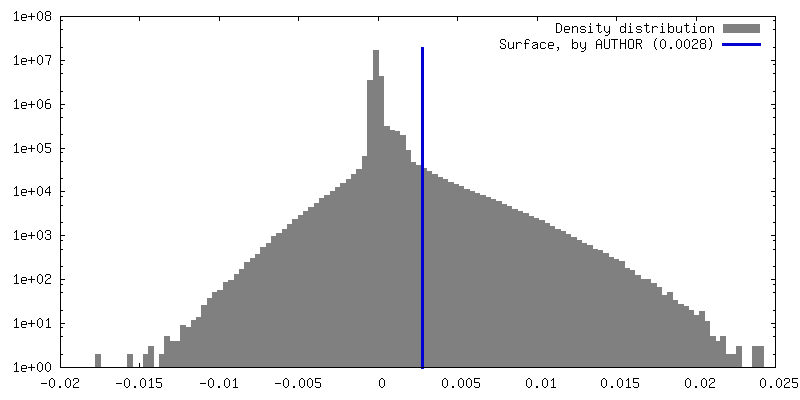
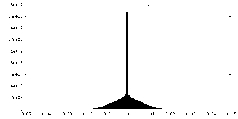
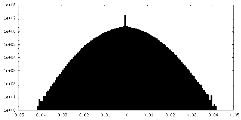
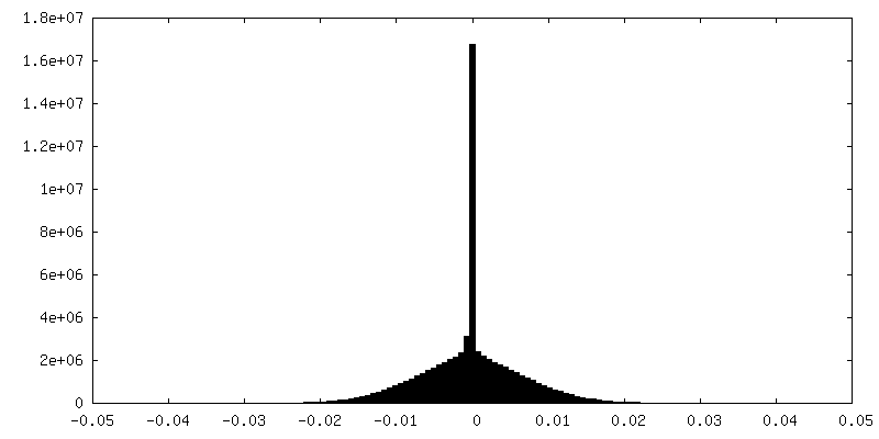
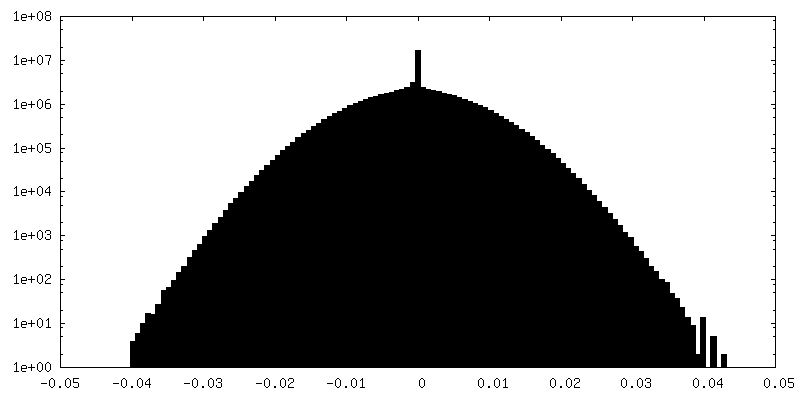
| Density Histograms |


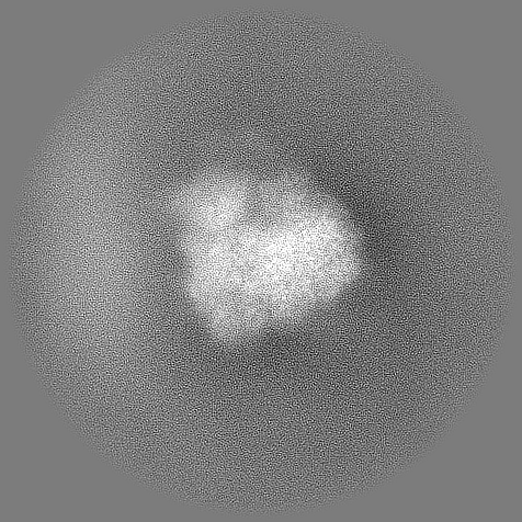
-Half map: From WARP/M
| File | emd_16537_half_map_1.map | ||||||||||||
|---|---|---|---|---|---|---|---|---|---|---|---|---|---|
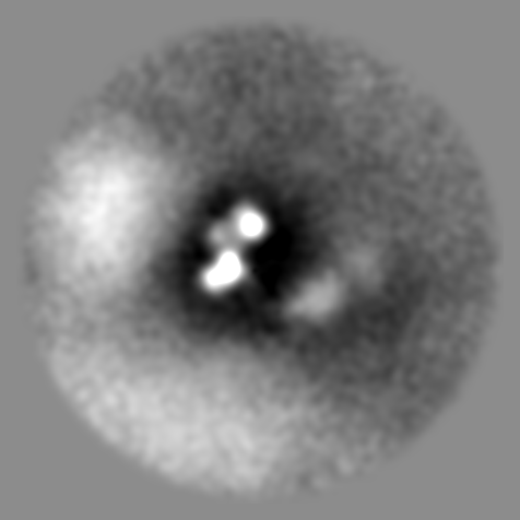
| Annotation | From WARP/M | ||||||||||||
| Projections & Slices |
| ||||||||||||
| Density Histograms |

-Half map: From WARP/M
| File | emd_16537_half_map_2.map | ||||||||||||
|---|---|---|---|---|---|---|---|---|---|---|---|---|---|
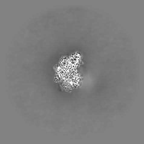
| Annotation | From WARP/M | ||||||||||||
| Projections & Slices |
| ||||||||||||
| Density Histograms |

- Sample components
-Entire : S.Cerevisiae 80s ribosome large subunit
| Entire | Name: S.Cerevisiae 80s ribosome large subunit |
|---|---|
| Components |
|
-Supramolecule #1: S.Cerevisiae 80s ribosome large subunit
| Supramolecule | Name: S.Cerevisiae 80s ribosome large subunit / type: complex / ID: 1 / Parent: 0 |
|---|---|
| Source (natural) | Organism: |
| Molecular weight | Theoretical: 3.5 MDa |
-Experimental details
-Structure determination
| Method | cryo EM |
|---|---|
 Processing Processing | subtomogram averaging |
| Aggregation state | cell |
-Sample preparation
| Buffer | pH: 7.5 |
|---|---|
| Vitrification | Cryogen name: ETHANE-PROPANE |
- Electron microscopy
| Microscope | FEI TITAN KRIOS |
|---|---|
| Image recording | Film or detector model: FEI FALCON IV (4k x 4k) / Average electron dose: 3.5 e/Å2 |
| Electron beam | Acceleration voltage: 300 kV / Electron source:  FIELD EMISSION GUN FIELD EMISSION GUN |
| Electron optics | Illumination mode: FLOOD BEAM / Imaging mode: BRIGHT FIELD / Nominal defocus max: 3.5 µm / Nominal defocus min: 1.5 µm |
| Experimental equipment |  Model: Titan Krios / Image courtesy: FEI Company |
-Image processing
| Final reconstruction | Applied symmetry - Point group: C1 (asymmetric) / Resolution.type: BY AUTHOR / Resolution: 4.6 Å / Resolution method: FSC 0.143 CUT-OFF / Number subtomograms used: 12500 |
|---|---|
| Extraction | Number tomograms: 91 / Number images used: 45500 |
| Final angle assignment | Type: MAXIMUM LIKELIHOOD |
