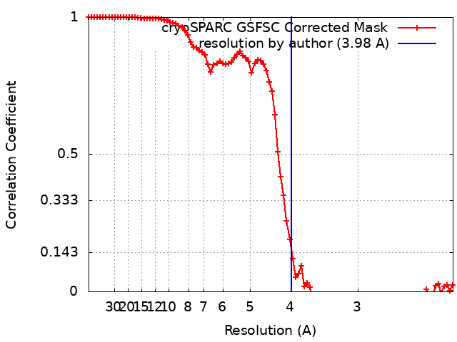
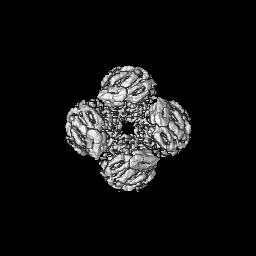
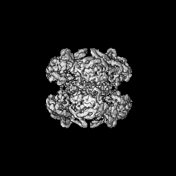
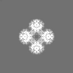
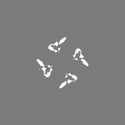
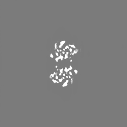
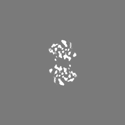
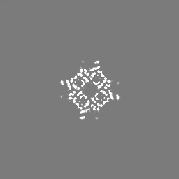
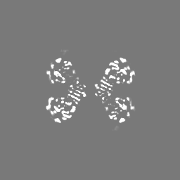
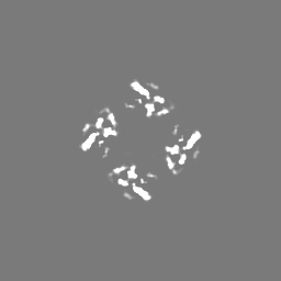
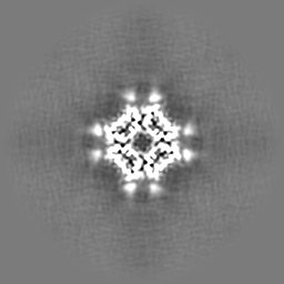
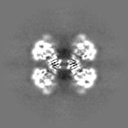
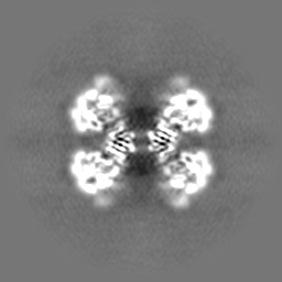
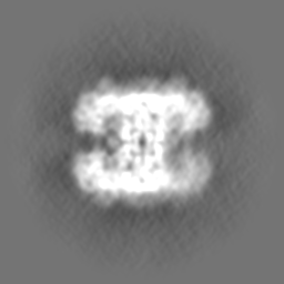
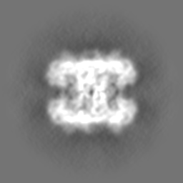
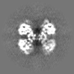
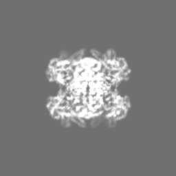
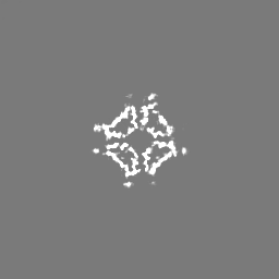
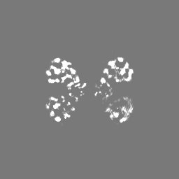
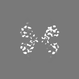
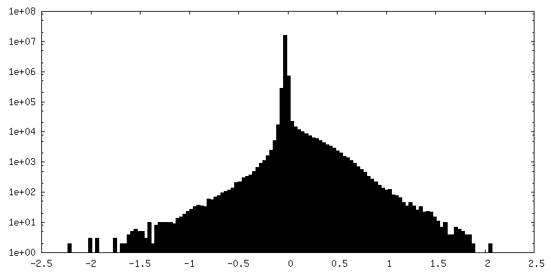
Journal: mBio / Year: 2023 Title: CryoEM analysis of the essential native UDP-glucose pyrophosphorylase from reveals key conformations for activity regulation and function. Authors: Xu Han / Cecilia D'Angelo / Ainara Otamendi / Javier O Cifuente / Elisa de Astigarraga / Borja Ochoa-Lizarralde / Martin Grininger / Francoise H Routier / Marcelo E Guerin / Jana Fuehring / ...Authors: Xu Han / Cecilia D'Angelo / Ainara Otamendi / Javier O Cifuente / Elisa de Astigarraga / Borja Ochoa-Lizarralde / Martin Grininger / Francoise H Routier / Marcelo E Guerin / Jana Fuehring / Oier Etxebeste / Sean R Connell / Abstract: Invasive aspergillosis is one of the most serious clinical invasive fungal infections, resulting in a high case fatality rate among immunocompromised patients. The disease is caused by saprophytic ...Invasive aspergillosis is one of the most serious clinical invasive fungal infections, resulting in a high case fatality rate among immunocompromised patients. The disease is caused by saprophytic molds in the genus , including , the most significant pathogenic species. The fungal cell wall, an essential structure mainly composed of glucan, chitin, galactomannan, and galactosaminogalactan, represents an important target for the development of antifungal drugs. UDP (uridine diphosphate)-glucose pyrophosphorylase (UGP) is a central enzyme in the metabolism of carbohydrates that catalyzes the biosynthesis of UDP-glucose, a key precursor of fungal cell wall polysaccharides. Here, we demonstrate that the function of UGP is vital for (UGP). To understand the molecular basis of UGP function, we describe a cryoEM structure (global resolution of 3.5 Å for the locally refined subunit and 4 Å for the octameric complex) of a native UGP. The structure reveals an octameric architecture with each subunit comprising an N-terminal α-helical domain, a central catalytic glycosyltransferase A-like (GT-A-like) domain, and a C-terminal (CT) left-handed β-helix oligomerization domain. UGP displays unprecedented conformational variability between the CT oligomerization domain and the central GT-A-like catalytic domain. In combination with activity measurements and bioinformatics analysis, we unveil the molecular mechanism of substrate recognition and specificity for UGP. Altogether, our study not only contributes to understanding the molecular mechanism of catalysis/regulation of an important class of enzymes but also provides the genetic, biochemical, and structural groundwork for the future exploitation of UGP as a potential antifungal target. IMPORTANCE Fungi cause diverse diseases in humans, ranging from allergic syndromes to life-threatening invasive diseases, together affecting more than a billion people worldwide. Increasing drug resistance in species represents an emerging global health threat, making the design of antifungals with novel mechanisms of action a worldwide priority. The cryoEM structure of UDP (uridine diphosphate)-glucose pyrophosphorylase (UGP) from the filamentous fungus reveals an octameric architecture displaying unprecedented conformational variability between the C-terminal oligomerization domain and the central glycosyltransferase A-like catalytic domain in the individual protomers. While the active site and oligomerization interfaces are more highly conserved, these dynamic interfaces include motifs restricted to specific clades of filamentous fungi. Functional study of these motifs could lead to the definition of new targets for antifungals inhibiting UGP activity and, thus, the architecture of the cell wall of filamentous fungal pathogens.
In the structure databanks used in Yorodumi, some data are registered as the other names, "COVID-19 virus" and "2019-nCoV". Here are the details of the virus and the list of structure data.
Jan 31, 2019. EMDB accession codes are about to change! (news from PDBe EMDB page)
EMDB accession codes are about to change! (news from PDBe EMDB page)
The allocation of 4 digits for EMDB accession codes will soon come to an end. Whilst these codes will remain in use, new EMDB accession codes will include an additional digit and will expand incrementally as the available range of codes is exhausted. The current 4-digit format prefixed with “EMD-” (i.e. EMD-XXXX) will advance to a 5-digit format (i.e. EMD-XXXXX), and so on. It is currently estimated that the 4-digit codes will be depleted around Spring 2019, at which point the 5-digit format will come into force.
The EM Navigator/Yorodumi systems omit the EMD- prefix.
Related info.:Q: What is EMD? / ID/Accession-code notation in Yorodumi/EM Navigator
Yorodumi is a browser for structure data from EMDB, PDB, SASBDB, etc.
This page is also the successor to EM Navigator detail page, and also detail information page/front-end page for Omokage search.
The word "yorodu" (or yorozu) is an old Japanese word meaning "ten thousand". "mi" (miru) is to see.
Related info.:EMDB / PDB / SASBDB / Comparison of 3 databanks / Yorodumi Search / Aug 31, 2016. New EM Navigator & Yorodumi / Yorodumi Papers / Jmol/JSmol / Function and homology information / Changes in new EM Navigator and Yorodumi
 Movie
Movie Controller

 Yorodumi
 Open data
 Basic information
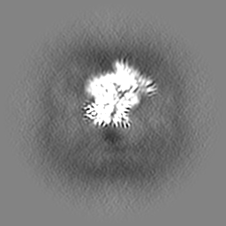
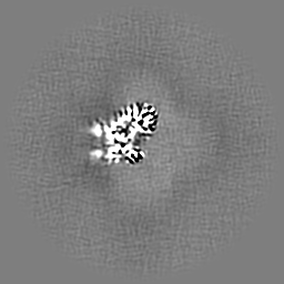
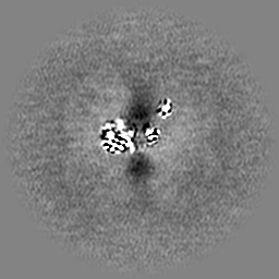
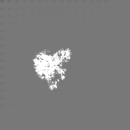
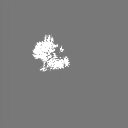
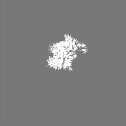
 Map data
 Sample
 Keywords
 Function and homology information
 Authors
 Spain, 1 items
  Citation
 Structure visualization
 Downloads & links
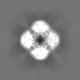
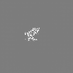
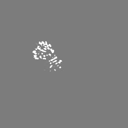
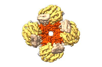 emd_16357.png
 http://ftp.pdbj.org/pub/emdb/structures/EMD-16357


 Z (Sec.)
 Y (Row.)
 X (Col.)


 Sample components
 Processing
 Electron microscopy
 FIELD EMISSION GUN