 Movie
Movie Controller

[English] 日本語
 Yorodumi
- EMDB-15547: In situ cryo-electron tomogram of a bulk autophagy autophagosome ... -
+ Open data

- Basic information
| Entry |  | |||||||||
|---|---|---|---|---|---|---|---|---|---|---|
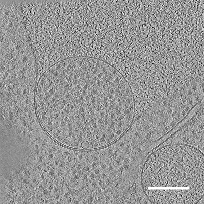
| Title | In situ cryo-electron tomogram of a bulk autophagy autophagosome fusing with the vacuole in S. cerevisiae #1 | |||||||||
 Map data Map data | Fusion of autophagosome with the vacuole, in situ tomogram in S. cerevisiae | |||||||||
 Sample Sample |
| |||||||||
 Keywords Keywords | Autophagy / phagophore / fusion / yeast / bulk / degradation / RIBOSOME | |||||||||
| Biological species |  | |||||||||
| Method | electron tomography / cryo EM | |||||||||
 Authors Authors | Bieber A / Capitanio C / Erdmann PS / Schulman BA / Baumeister W / Wilfling F | |||||||||
| Funding support |  United States, 1 items United States, 1 items
| |||||||||
 Citation Citation | Journal: Proc Natl Acad Sci U S A / Year: 2022 Title: In situ structural analysis reveals membrane shape transitions during autophagosome formation. Authors: Anna Bieber / Cristina Capitanio / Philipp S Erdmann / Fabian Fiedler / Florian Beck / Chia-Wei Lee / Delong Li / Gerhard Hummer / Brenda A Schulman / Wolfgang Baumeister / Florian Wilfling /   Abstract: Autophagosomes are unique organelles that form de novo as double-membrane vesicles engulfing cytosolic material for destruction. Their biogenesis involves membrane transformations of distinctly ...Autophagosomes are unique organelles that form de novo as double-membrane vesicles engulfing cytosolic material for destruction. Their biogenesis involves membrane transformations of distinctly shaped intermediates whose ultrastructure is poorly understood. Here, we combine cell biology, correlative cryo-electron tomography (cryo-ET), and extensive data analysis to reveal the step-by-step structural progression of autophagosome biogenesis at high resolution directly within yeast cells. The analysis uncovers an unexpectedly thin intermembrane distance that is dilated at the phagophore rim. Mapping of individual autophagic structures onto a timeline based on geometric features reveals a dynamical change of membrane shape and curvature in growing phagophores. Moreover, our tomograms show the organelle interactome of growing autophagosomes, highlighting a polar organization of contact sites between the phagophore and organelles, such as the vacuole and the endoplasmic reticulum (ER). Collectively, these findings have important implications for the contribution of different membrane sources during autophagy and for the forces shaping and driving phagophores toward closure without a templating cargo. | |||||||||
| History |
|
- Structure visualization
| Supplemental images |
|---|
- Downloads & links
-EMDB archive
| Map data | emd_15547.map.gz | 761.3 MB |  EMDB map data format EMDB map data format | |
|---|---|---|---|---|
| Header (meta data) | emd-15547-v30.xmlemd-15547.xml | 11.4 KB 11.4 KB | Display Display | EMDB header |
| Images |  emd_15547.png emd_15547.png | 265.3 KB | ||
| Filedesc metadata | emd-15547.cif.gz | 4.1 KB | ||
| Archive directory |  http://ftp.pdbj.org/pub/emdb/structures/EMD-15547ftp://ftp.pdbj.org/pub/emdb/structures/EMD-15547 http://ftp.pdbj.org/pub/emdb/structures/EMD-15547ftp://ftp.pdbj.org/pub/emdb/structures/EMD-15547 | HTTPS FTP |
-Related structure data


-Links
| EMDB pages | EMDB (EBI/PDBe) / EMDataResource |
|---|
-Map
| File | Download / File: emd_15547.map.gz / Format: CCP4 / Size: 821.3 MB / Type: IMAGE STORED AS FLOATING POINT NUMBER (4 BYTES) | ||||||||||||||||||||||||||||||||
|---|---|---|---|---|---|---|---|---|---|---|---|---|---|---|---|---|---|---|---|---|---|---|---|---|---|---|---|---|---|---|---|---|---|
| Annotation | Fusion of autophagosome with the vacuole, in situ tomogram in S. cerevisiae | ||||||||||||||||||||||||||||||||
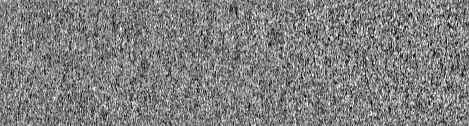
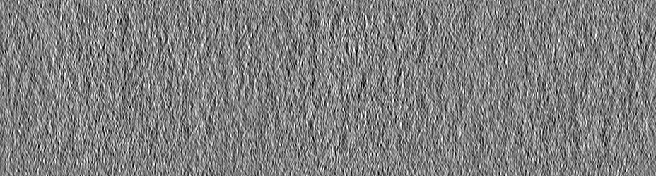
| Projections & slices | Image control
Images are generated by Spider. generated in cubic-lattice coordinate | ||||||||||||||||||||||||||||||||
| Voxel size | X=Y=Z: 14.08 Å | ||||||||||||||||||||||||||||||||
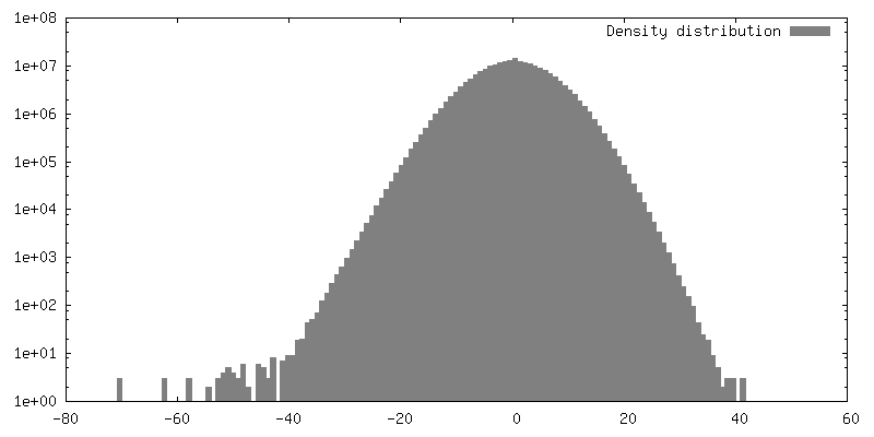
| Density |
| ||||||||||||||||||||||||||||||||
| Symmetry | Space group: 1 | ||||||||||||||||||||||||||||||||
| Details | EMDB XML:
|
 Z (Sec.)
 Y (Row.)
 X (Col.)


-Supplemental data
- Sample components
-Entire : In situ cryo-electron tomogram of a bulk autophagy autophagosome ...
| Entire | Name: In situ cryo-electron tomogram of a bulk autophagy autophagosome fusing with the vacuole in S. cerevisiae #1 |
|---|---|
| Components |
|
-Supramolecule #1: In situ cryo-electron tomogram of a bulk autophagy autophagosome ...
| Supramolecule | Name: In situ cryo-electron tomogram of a bulk autophagy autophagosome fusing with the vacuole in S. cerevisiae #1 type: cell / ID: 1 / Parent: 0 Details: Autophagosome fusing with the vacuole. The position of the autophagosome corresponds to mCherry-Atg8 signal detected by cryo-fluorescence microscopy. |
|---|---|
| Source (natural) | Organism: |
-Experimental details
-Structure determination
| Method | cryo EM |
|---|---|
 Processing Processing | electron tomography |
| Aggregation state | cell |
-Sample preparation
| Buffer | pH: 7 |
|---|---|
| Grid | Model: Quantifoil / Support film - topology: HOLEY |
| Vitrification | Cryogen name: ETHANE-PROPANE / Chamber temperature: 295 K / Instrument: FEI VITROBOT MARK IV |
| Details | S. cerevisiae. 2 hours starvation in SD-N. |
| Sectioning | Focused ion beam - Instrument: OTHER / Focused ion beam - Ion: OTHER / Focused ion beam - Voltage: 30 / Focused ion beam - Current: 0.3 / Focused ion beam - Duration: 2400 / Focused ion beam - Temperature: 91 K / Focused ion beam - Initial thickness: 7000 / Focused ion beam - Final thickness: 150 Focused ion beam - Details: Correlative FIB-milling. The lamella was prepared in the location of fluorescence signal.. The value given for _em_focused_ion_beam.instrument is Other. This is not in a ...Focused ion beam - Details: Correlative FIB-milling. The lamella was prepared in the location of fluorescence signal.. The value given for _em_focused_ion_beam.instrument is Other. This is not in a list of allowed values {'DB235', 'OTHER'} so OTHER is written into the XML file. |
- Electron microscopy
| Microscope | FEI TITAN KRIOS |
|---|---|
| Image recording | Film or detector model: GATAN K2 SUMMIT (4k x 4k) / Detector mode: COUNTING / Digitization - Dimensions - Width: 3712 pixel / Digitization - Dimensions - Height: 3712 pixel / Number real images: 60 / Average exposure time: 2.0 sec. / Average electron dose: 120.0 e/Å2 |
| Electron beam | Acceleration voltage: 300 kV / Electron source:  FIELD EMISSION GUN FIELD EMISSION GUN |
| Electron optics | Illumination mode: FLOOD BEAM / Imaging mode: BRIGHT FIELD / Nominal defocus max: 4.5 µm / Nominal defocus min: 4.0 µm / Nominal magnification: 42000 |
| Sample stage | Specimen holder model: FEI TITAN KRIOS AUTOGRID HOLDER / Cooling holder cryogen: NITROGEN |
| Experimental equipment |  Model: Titan Krios / Image courtesy: FEI Company |
-Image processing
| Final reconstruction | Software - Name: IMOD / Number images used: 55 |
|---|
