- EMDB-15394: Cell-cell contact between two PTK-1 cells -
+
データを開く
IDまたはキーワード:
読み込み中...
-
基本情報
登録情報
データベース: EMDB / ID: EMD-15394
タイトル
Cell-cell contact between two PTK-1 cells
マップデータ
Raw tomographic reconstruction of a cryo-FIB lamella through a cell-cell contact of two PTK-1 cells. "Contour level" is a placeholder. Use "Auto" button in IMOD.
ジャーナル: Nat Commun / 年: 2022 タイトル: Morphological control enables nanometer-scale dissection of cell-cell signaling complexes. 著者: Liam P Dow / Guido Gaietta / Yair Kaufman / Mark F Swift / Moara Lemos / Kerry Lane / Matthew Hopcroft / Armel Bezault / Cécile Sauvanet / Niels Volkmann / Beth L Pruitt / Dorit Hanein / 要旨: Protein micropatterning enables robust control of cell positioning on electron-microscopy substrates for cryogenic electron tomography (cryo-ET). However, the combination of regulated cell boundaries ...Protein micropatterning enables robust control of cell positioning on electron-microscopy substrates for cryogenic electron tomography (cryo-ET). However, the combination of regulated cell boundaries and the underlying electron-microscopy substrate (EM-grids) provides a poorly understood microenvironment for cell biology. Because substrate stiffness and morphology affect cellular behavior, we devised protocols to characterize the nanometer-scale details of the protein micropatterns on EM-grids by combining cryo-ET, atomic force microscopy, and scanning electron microscopy. Measuring force displacement characteristics of holey carbon EM-grids, we found that their effective spring constant is similar to physiological values expected from skin tissues. Despite their apparent smoothness at light-microscopy resolution, spatial boundaries of the protein micropatterns are irregular at nanometer scale. Our protein micropatterning workflow provides the means to steer both positioning and morphology of cell doublets to determine nanometer details of punctate adherens junctions. Our workflow serves as the foundation for studying the fundamental structural changes governing cell-cell signaling.
ダウンロード / ファイル: emd_15394.map.gz / 形式: CCP4 / 大きさ: 928 MB / タイプ: IMAGE STORED AS SIGNED BYTE
注釈
Raw tomographic reconstruction of a cryo-FIB lamella through a cell-cell contact of two PTK-1 cells. "Contour level" is a placeholder. Use "Auto" button in IMOD.
ボクセルのサイズ
X=Y=Z: 8.114 Å
密度
最小 - 最大
-128.0 - 127.0
平均 (標準偏差)
6.7312713 (±15.179936)
対称性
空間群: 1
詳細
EMDB XML:
マップ形状
Axis order
X
Y
Z
Origin
0
0
89
サイズ
2048
2048
232
Spacing
2048
2048
232
セル
A: 16617.473 Å / B: 16617.473 Å / C: 1882.4481 Å α=β=γ: 90.0 °
-
添付データ
-
試料の構成要素
-
全体 : cell-cell contact
全体
名称: cell-cell contact
要素
細胞: cell-cell contact
-
超分子 #1: cell-cell contact
超分子
名称: cell-cell contact / タイプ: cell / ID: 1 / 親要素: 0 / 詳細: cell-cell contact between two PTK-1 cells
由来(天然)
生物種: Potorous tridactylus (ハナナガネズミカンガルー)
-
実験情報
-
構造解析
手法
クライオ電子顕微鏡法
解析
電子線トモグラフィー法
試料の集合状態
cell
-
試料調製
緩衝液
pH: 6.9
凍結
凍結剤: ETHANE
切片作成
その他: NO SECTIONING
-
電子顕微鏡法
顕微鏡
TFS GLACIOS
撮影
フィルム・検出器のモデル: FEI FALCON III (4k x 4k) 平均電子線量: 3.0 e/Å2
 ムービー
ムービー コントローラー


 データを開く
 基本情報
基本情報
 マップデータ
 試料
試料 Potorous tridactylus (ハナナガネズミカンガルー)
 データ登録者
 引用
引用

 構造の表示
構造の表示
 ダウンロードとリンク
 EMDBマップデータ形式
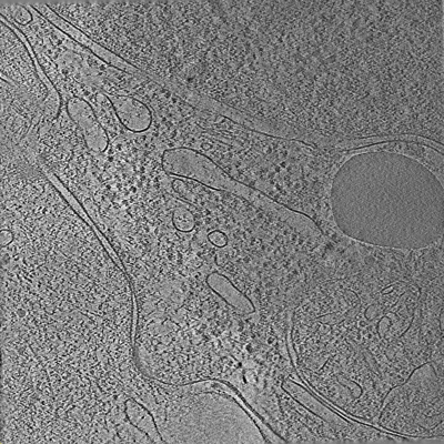 emd_15394.png
 http://ftp.pdbj.org/pub/emdb/structures/EMD-15394
 試料の構成要素
 解析
解析 電子顕微鏡法
 FIELD EMISSION GUN