- EMDB-15394: Cell-cell contact between two PTK-1 cells -
+
Open data
ID or keywords:
Loading...
-
Basic information
Entry
Database: EMDB / ID: EMD-15394
Title
Cell-cell contact between two PTK-1 cells
Map data
Raw tomographic reconstruction of a cryo-FIB lamella through a cell-cell contact of two PTK-1 cells. "Contour level" is a placeholder. Use "Auto" button in IMOD.
Journal: Nat Commun / Year: 2022 Title: Morphological control enables nanometer-scale dissection of cell-cell signaling complexes. Authors: Liam P Dow / Guido Gaietta / Yair Kaufman / Mark F Swift / Moara Lemos / Kerry Lane / Matthew Hopcroft / Armel Bezault / Cécile Sauvanet / Niels Volkmann / Beth L Pruitt / Dorit Hanein / Abstract: Protein micropatterning enables robust control of cell positioning on electron-microscopy substrates for cryogenic electron tomography (cryo-ET). However, the combination of regulated cell boundaries ...Protein micropatterning enables robust control of cell positioning on electron-microscopy substrates for cryogenic electron tomography (cryo-ET). However, the combination of regulated cell boundaries and the underlying electron-microscopy substrate (EM-grids) provides a poorly understood microenvironment for cell biology. Because substrate stiffness and morphology affect cellular behavior, we devised protocols to characterize the nanometer-scale details of the protein micropatterns on EM-grids by combining cryo-ET, atomic force microscopy, and scanning electron microscopy. Measuring force displacement characteristics of holey carbon EM-grids, we found that their effective spring constant is similar to physiological values expected from skin tissues. Despite their apparent smoothness at light-microscopy resolution, spatial boundaries of the protein micropatterns are irregular at nanometer scale. Our protein micropatterning workflow provides the means to steer both positioning and morphology of cell doublets to determine nanometer details of punctate adherens junctions. Our workflow serves as the foundation for studying the fundamental structural changes governing cell-cell signaling.
Download / File: emd_15394.map.gz / Format: CCP4 / Size: 928 MB / Type: IMAGE STORED AS SIGNED BYTE
Annotation
Raw tomographic reconstruction of a cryo-FIB lamella through a cell-cell contact of two PTK-1 cells. "Contour level" is a placeholder. Use "Auto" button in IMOD.
In the structure databanks used in Yorodumi, some data are registered as the other names, "COVID-19 virus" and "2019-nCoV". Here are the details of the virus and the list of structure data.
Jan 31, 2019. EMDB accession codes are about to change! (news from PDBe EMDB page)
EMDB accession codes are about to change! (news from PDBe EMDB page)
The allocation of 4 digits for EMDB accession codes will soon come to an end. Whilst these codes will remain in use, new EMDB accession codes will include an additional digit and will expand incrementally as the available range of codes is exhausted. The current 4-digit format prefixed with “EMD-” (i.e. EMD-XXXX) will advance to a 5-digit format (i.e. EMD-XXXXX), and so on. It is currently estimated that the 4-digit codes will be depleted around Spring 2019, at which point the 5-digit format will come into force.
The EM Navigator/Yorodumi systems omit the EMD- prefix.
Related info.:Q: What is EMD? / ID/Accession-code notation in Yorodumi/EM Navigator
Yorodumi is a browser for structure data from EMDB, PDB, SASBDB, etc.
This page is also the successor to EM Navigator detail page, and also detail information page/front-end page for Omokage search.
The word "yorodu" (or yorozu) is an old Japanese word meaning "ten thousand". "mi" (miru) is to see.
Related info.:EMDB / PDB / SASBDB / Comparison of 3 databanks / Yorodumi Search / Aug 31, 2016. New EM Navigator & Yorodumi / Yorodumi Papers / Jmol/JSmol / Function and homology information / Changes in new EM Navigator and Yorodumi
 Movie
Movie Controller


 Open data
 Basic information
 Map data
 Sample
 Potorous tridactylus (long-nosed potoroo)
 Authors
 Citation

 Structure visualization
 Downloads & links
 EMDB map data format
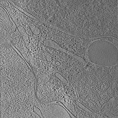 emd_15394.png
 http://ftp.pdbj.org/pub/emdb/structures/EMD-15394
 Z (Sec.)
 Y (Row.)
 X (Col.)


 Sample components
 Processing
 Electron microscopy
 FIELD EMISSION GUN