National Institutes of Health/National Institute of Neurological Disorders and Stroke (NIH/NINDS)
United States
Citation
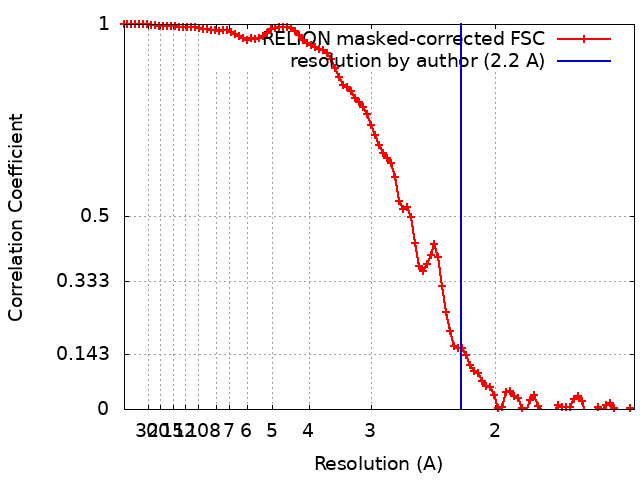
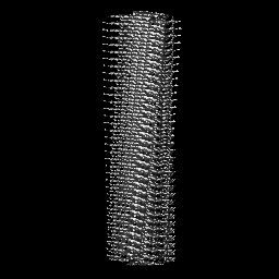
Journal: Nature / Year: 2022 Title: Structures of α-synuclein filaments from human brains with Lewy pathology. Authors: Yang Yang / Yang Shi / Manuel Schweighauser / Xianjun Zhang / Abhay Kotecha / Alexey G Murzin / Holly J Garringer / Patrick W Cullinane / Yuko Saito / Tatiana Foroud / Thomas T Warner / ...Authors: Yang Yang / Yang Shi / Manuel Schweighauser / Xianjun Zhang / Abhay Kotecha / Alexey G Murzin / Holly J Garringer / Patrick W Cullinane / Yuko Saito / Tatiana Foroud / Thomas T Warner / Kazuko Hasegawa / Ruben Vidal / Shigeo Murayama / Tamas Revesz / Bernardino Ghetti / Masato Hasegawa / Tammaryn Lashley / Sjors H W Scheres / Michel Goedert / Abstract: Parkinson's disease (PD) is the most common movement disorder, with resting tremor, rigidity, bradykinesia and postural instability being major symptoms. Neuropathologically, it is characterized by ...Parkinson's disease (PD) is the most common movement disorder, with resting tremor, rigidity, bradykinesia and postural instability being major symptoms. Neuropathologically, it is characterized by the presence of abundant filamentous inclusions of α-synuclein in the form of Lewy bodies and Lewy neurites in some brain cells, including dopaminergic nerve cells of the substantia nigra. PD is increasingly recognised as a multisystem disorder, with cognitive decline being one of its most common non-motor symptoms. Many patients with PD develop dementia more than 10 years after diagnosis. PD dementia (PDD) is clinically and neuropathologically similar to dementia with Lewy bodies (DLB), which is diagnosed when cognitive impairment precedes parkinsonian motor signs or begins within one year from their onset. In PDD, cognitive impairment develops in the setting of well-established PD. Besides PD and DLB, multiple system atrophy (MSA) is the third major synucleinopathy. It is characterized by the presence of abundant filamentous α-synuclein inclusions in brain cells, especially oligodendrocytes (Papp-Lantos bodies). We previously reported the electron cryo-microscopy structures of two types of α-synuclein filament extracted from the brains of individuals with MSA. Each filament type is made of two different protofilaments. Here we report that the cryo-electron microscopy structures of α-synuclein filaments from the brains of individuals with PD, PDD and DLB are made of a single protofilament (Lewy fold) that is markedly different from the protofilaments of MSA. These findings establish the existence of distinct molecular conformers of assembled α-synuclein in neurodegenerative disease.
In the structure databanks used in Yorodumi, some data are registered as the other names, "COVID-19 virus" and "2019-nCoV". Here are the details of the virus and the list of structure data.
Jan 31, 2019. EMDB accession codes are about to change! (news from PDBe EMDB page)
EMDB accession codes are about to change! (news from PDBe EMDB page)
The allocation of 4 digits for EMDB accession codes will soon come to an end. Whilst these codes will remain in use, new EMDB accession codes will include an additional digit and will expand incrementally as the available range of codes is exhausted. The current 4-digit format prefixed with “EMD-” (i.e. EMD-XXXX) will advance to a 5-digit format (i.e. EMD-XXXXX), and so on. It is currently estimated that the 4-digit codes will be depleted around Spring 2019, at which point the 5-digit format will come into force.
The EM Navigator/Yorodumi systems omit the EMD- prefix.
Related info.:Q: What is EMD? / ID/Accession-code notation in Yorodumi/EM Navigator
Yorodumi is a browser for structure data from EMDB, PDB, SASBDB, etc.
This page is also the successor to EM Navigator detail page, and also detail information page/front-end page for Omokage search.
The word "yorodu" (or yorozu) is an old Japanese word meaning "ten thousand". "mi" (miru) is to see.
Related info.:EMDB / PDB / SASBDB / Comparison of 3 databanks / Yorodumi Search / Aug 31, 2016. New EM Navigator & Yorodumi / Yorodumi Papers / Jmol/JSmol / Function and homology information / Changes in new EM Navigator and Yorodumi
 Movie
Movie Controller

 Yorodumi
 Open data
 Basic information
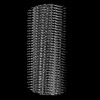
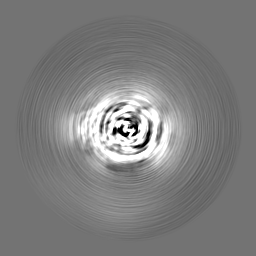
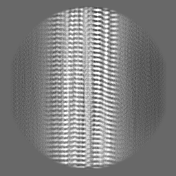
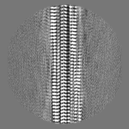
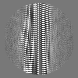
 Map data
 Sample
 Keywords
 Function and homology information
 Homo sapiens (human)
 Authors
 United Kingdom,
  United States, 4 items
  Citation

 Structure visualization
 Downloads & links
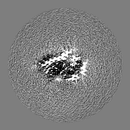
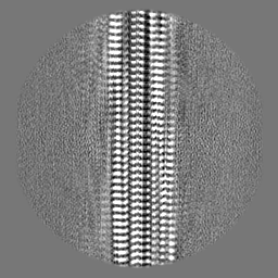
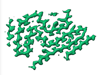 emd_15285.png
 http://ftp.pdbj.org/pub/emdb/structures/EMD-15285
 Z (Sec.)
 Y (Row.)
 X (Col.)


 Sample components
 Processing
 Electron microscopy
 FIELD EMISSION GUN