 Movie
Movie Controller


+ Open data

- Basic information
| Entry | 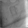 | ||||||||||||||||||||||||
|---|---|---|---|---|---|---|---|---|---|---|---|---|---|---|---|---|---|---|---|---|---|---|---|---|---|
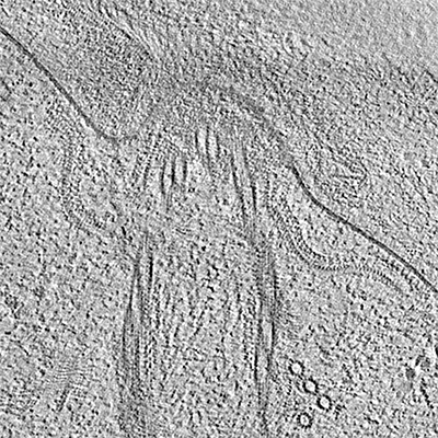
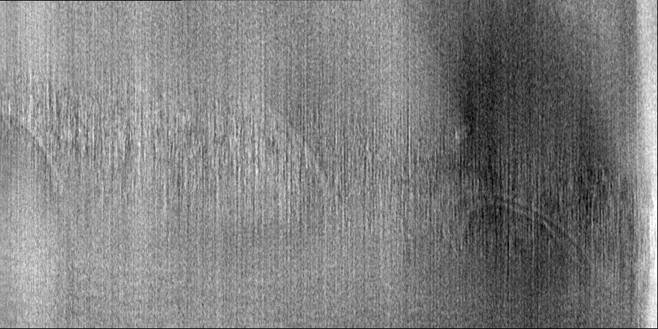
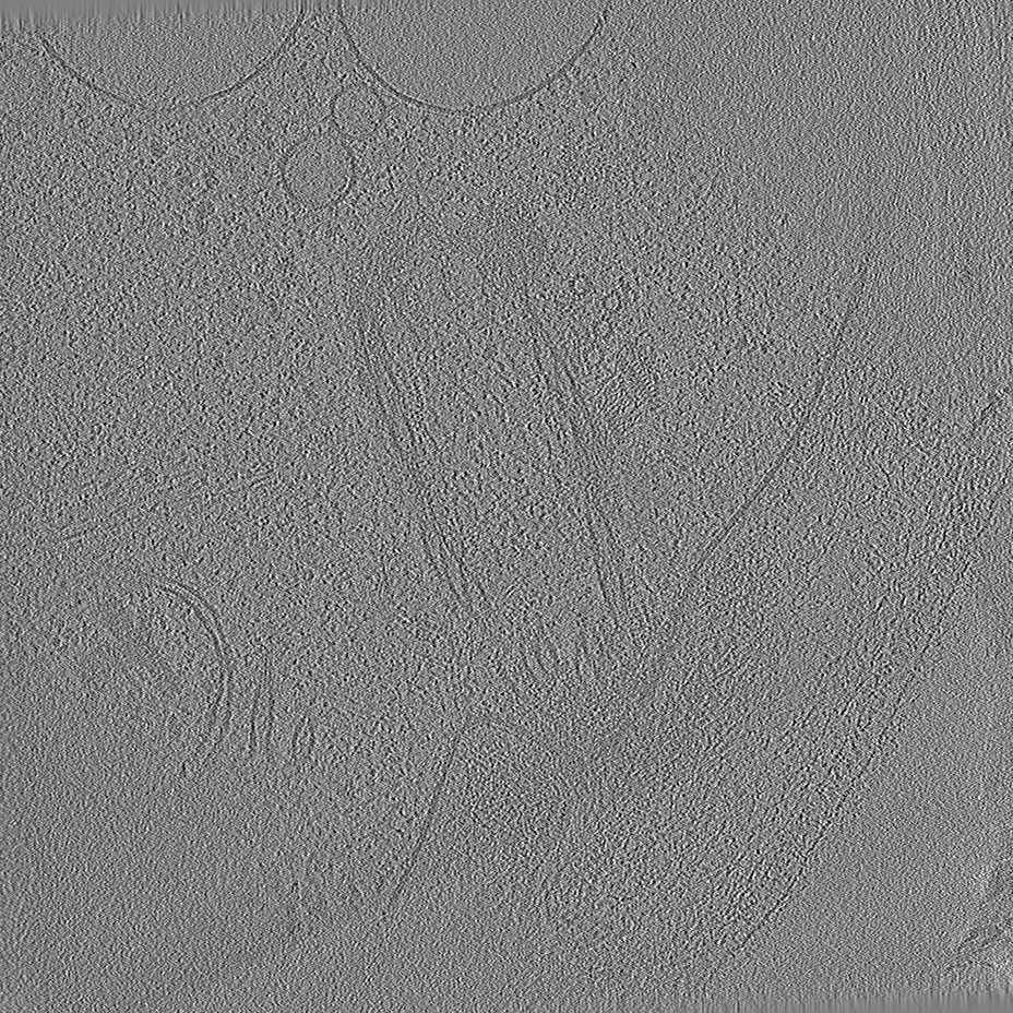
| Title | Tomogram #3 of the C. reinhardtii ciliary transition zone | ||||||||||||||||||||||||
 Map data Map data | Tomogram at bin4 deconvoluted using ad-hoc deconvolution filter (tom_deconv.m) | ||||||||||||||||||||||||
 Sample Sample |
| ||||||||||||||||||||||||
 Keywords Keywords | cilia / transition zone / flagella / ciliary pore / intraflagellar transport / motility / signaling / basal body / microtubules / chlamydomonas / STRUCTURAL PROTEIN | ||||||||||||||||||||||||
| Biological species |   Chlamydomonas reinhardtii (plant) Chlamydomonas reinhardtii (plant) | ||||||||||||||||||||||||
| Method | electron tomography / cryo EM | ||||||||||||||||||||||||
 Authors Authors | van den Hoek HG / Schaffer M / Erdmann PS / Wan WN / Plitzko JM / Baumeister W / Engel BD | ||||||||||||||||||||||||
| Funding support | European Union,  Germany, Germany,  Switzerland, 7 items Switzerland, 7 items
| ||||||||||||||||||||||||
 Citation Citation | Journal: Science / Year: 2022 Title: In situ architecture of the ciliary base reveals the stepwise assembly of intraflagellar transport trains. Authors: Hugo van den Hoek / Nikolai Klena / Mareike A Jordan / Gonzalo Alvarez Viar / Ricardo D Righetto / Miroslava Schaffer / Philipp S Erdmann / William Wan / Stefan Geimer / Jürgen M Plitzko / ...Authors: Hugo van den Hoek / Nikolai Klena / Mareike A Jordan / Gonzalo Alvarez Viar / Ricardo D Righetto / Miroslava Schaffer / Philipp S Erdmann / William Wan / Stefan Geimer / Jürgen M Plitzko / Wolfgang Baumeister / Gaia Pigino / Virginie Hamel / Paul Guichard / Benjamin D Engel /   Abstract: The cilium is an antenna-like organelle that performs numerous cellular functions, including motility, sensing, and signaling. The base of the cilium contains a selective barrier that regulates the ...The cilium is an antenna-like organelle that performs numerous cellular functions, including motility, sensing, and signaling. The base of the cilium contains a selective barrier that regulates the entry of large intraflagellar transport (IFT) trains, which carry cargo proteins required for ciliary assembly and maintenance. However, the native architecture of the ciliary base and the process of IFT train assembly remain unresolved. In this work, we used in situ cryo-electron tomography to reveal native structures of the transition zone region and assembling IFT trains at the ciliary base in . We combined this direct cellular visualization with ultrastructure expansion microscopy to describe the front-to-back stepwise assembly of IFT trains: IFT-B forms the backbone, onto which bind IFT-A, dynein-1b, and finally kinesin-2 before entry into the cilium. | ||||||||||||||||||||||||
| History |
|
- Structure visualization
| Supplemental images |
|---|
- Downloads & links
-EMDB archive
| Map data | emd_15262.map.gz | 385.5 MB |  EMDB map data format EMDB map data format | |
|---|---|---|---|---|
| Header (meta data) | emd-15262-v30.xmlemd-15262.xml | 14.1 KB 14.1 KB | Display Display | EMDB header |
| Images |  emd_15262.png emd_15262.png | 295 KB | ||
| Filedesc metadata | emd-15262.cif.gz | 4.6 KB | ||
| Others | emd_15262_additional_1.map.gz | 380.2 MB | ||
| Archive directory |  http://ftp.pdbj.org/pub/emdb/structures/EMD-15262ftp://ftp.pdbj.org/pub/emdb/structures/EMD-15262 http://ftp.pdbj.org/pub/emdb/structures/EMD-15262ftp://ftp.pdbj.org/pub/emdb/structures/EMD-15262 | HTTPS FTP |
-Validation report
| Summary document | emd_15262_validation.pdf.gz | 470.1 KB | Display | EMDB validaton report |
|---|---|---|---|---|
| Full document | emd_15262_full_validation.pdf.gz | 469.7 KB | Display | |
| Data in XML | emd_15262_validation.xml.gz | 8 KB | Display | |
| Data in CIF | emd_15262_validation.cif.gz | 11 KB | Display | |
| Arichive directory | https://ftp.pdbj.org/pub/emdb/validation_reports/EMD-15262ftp://ftp.pdbj.org/pub/emdb/validation_reports/EMD-15262 | HTTPS FTP |
-Related structure data
| Related structure data | C: citing same article ( |
|---|---|
| EM raw data | EMPIAR-11078 (Title: In situ cryo-electron tomography of the C. reinhardtii ciliary transition zone Data size: 302.0 Data #1: Deconvoluted bin4 tomograms for visualization [reconstructed volumes] Data #2: CTF-corrected bin4 tomograms [reconstructed volumes] Data #3: CTF-corrected bin2 tomograms [reconstructed volumes] Data #4: Subtomograms at bin2 [subtomograms] / Data #5: Aligned tilt series stacks [tilt series] Data #6: Unaligned tilt series raw frames [micrographs - multiframe]) |


-Links
| EMDB pages | EMDB (EBI/PDBe) / EMDataResource |
|---|
-Map
| File | Download / File: emd_15262.map.gz / Format: CCP4 / Size: 762.2 MB / Type: IMAGE STORED AS SIGNED INTEGER (2 BYTES) | ||||||||||||||||||||||||||||||||
|---|---|---|---|---|---|---|---|---|---|---|---|---|---|---|---|---|---|---|---|---|---|---|---|---|---|---|---|---|---|---|---|---|---|
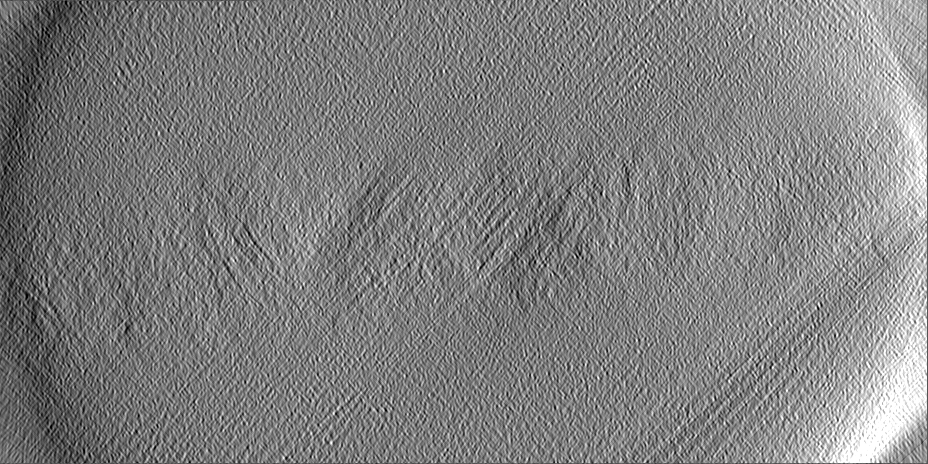
| Annotation | Tomogram at bin4 deconvoluted using ad-hoc deconvolution filter (tom_deconv.m) | ||||||||||||||||||||||||||||||||
| Projections & slices | Image control
Images are generated by Spider. generated in cubic-lattice coordinate | ||||||||||||||||||||||||||||||||
| Voxel size | X=Y=Z: 13.68 Å | ||||||||||||||||||||||||||||||||
| Density |
| ||||||||||||||||||||||||||||||||
| Symmetry | Space group: 1 | ||||||||||||||||||||||||||||||||
| Details | EMDB XML:
|
 Z (Sec.)
 Y (Row.)
 X (Col.)


-Supplemental data
-Additional map: Tomogram reconstructed with IMOD at bin4
| File | emd_15262_additional_1.map | ||||||||||||
|---|---|---|---|---|---|---|---|---|---|---|---|---|---|
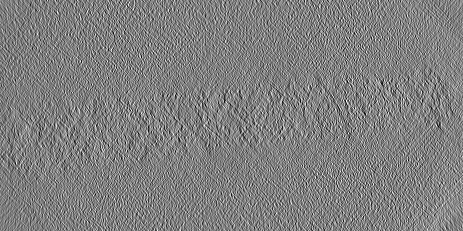
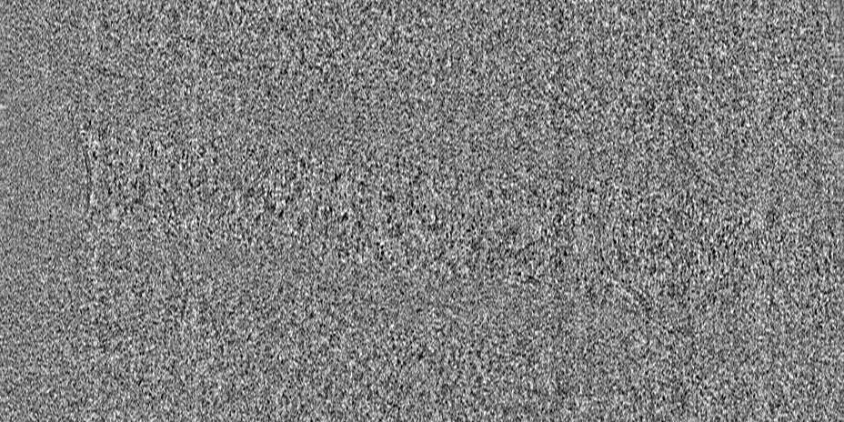
| Annotation | Tomogram reconstructed with IMOD at bin4 | ||||||||||||
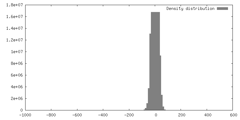
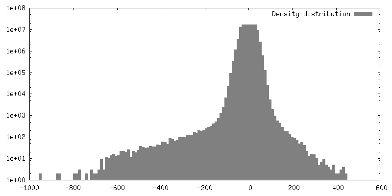
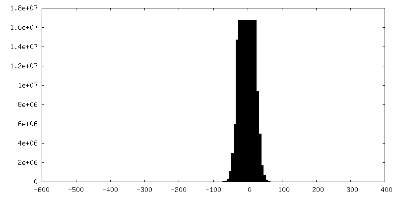
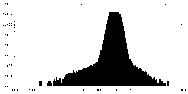
| Projections & Slices |
| ||||||||||||
| Density Histograms |

- Sample components
-Entire : C. reinhardtii ciliary transition zone
| Entire | Name: C. reinhardtii ciliary transition zone |
|---|---|
| Components |
|
-Supramolecule #1: C. reinhardtii ciliary transition zone
| Supramolecule | Name: C. reinhardtii ciliary transition zone / type: cell / ID: 1 / Parent: 0 |
|---|---|
| Source (natural) | Organism: Chlamydomonas reinhardtii (plant) / Strain: CC-3994 |
-Experimental details
-Structure determination
| Method | cryo EM |
|---|---|
 Processing Processing | electron tomography |
| Aggregation state | cell |
-Sample preparation
| Buffer | pH: 7 |
|---|---|
| Grid | Model: Quantifoil R2/1 / Material: COPPER / Support film - Material: CARBON / Support film - topology: CONTINUOUS / Pretreatment - Type: GLOW DISCHARGE |
| Vitrification | Cryogen name: ETHANE-PROPANE / Instrument: FEI VITROBOT MARK IV |
| Sectioning | Focused ion beam - Instrument: OTHER / Focused ion beam - Ion: OTHER / Focused ion beam - Voltage: 30 / Focused ion beam - Current: 0.03 / Focused ion beam - Duration: 2700 / Focused ion beam - Temperature: 91 K / Focused ion beam - Initial thickness: 6000 / Focused ion beam - Final thickness: 180 Focused ion beam - Details: The value given for _em_focused_ion_beam.instrument is FEI Quanta FIB. This is not in a list of allowed values {'DB235', 'OTHER'} so OTHER is written into the XML file. |
- Electron microscopy
| Microscope | FEI TITAN KRIOS |
|---|---|
| Specialist optics | Energy filter - Name: GIF Quantum LS / Energy filter - Slit width: 20 eV |
| Image recording | Film or detector model: GATAN K2 SUMMIT (4k x 4k) / Detector mode: COUNTING / Average electron dose: 100.0 e/Å2 |
| Electron beam | Acceleration voltage: 300 kV / Electron source:  FIELD EMISSION GUN FIELD EMISSION GUN |
| Electron optics | Illumination mode: FLOOD BEAM / Imaging mode: BRIGHT FIELD / Cs: 2.7 mm / Nominal defocus max: 6.0 µm / Nominal defocus min: 4.0 µm / Nominal magnification: 42000 |
| Sample stage | Specimen holder model: FEI TITAN KRIOS AUTOGRID HOLDER / Cooling holder cryogen: NITROGEN |
| Experimental equipment |  Model: Titan Krios / Image courtesy: FEI Company |
-Image processing
| Final reconstruction | Algorithm: BACK PROJECTION / Software - Name: STOPGAP (ver. 0.3) / Number images used: 60 |
|---|
