 Movie
Movie Controller


+ Open data

- Basic information
| Entry |  | |||||||||
|---|---|---|---|---|---|---|---|---|---|---|
| Title | Human PRPH2-ROM1 hetero-dimer | |||||||||
 Map data Map data | Non-sharpened map | |||||||||
 Sample Sample |
| |||||||||
 Keywords Keywords | PRPH2 / peripherin-2 / ROM1 / hetero-complex / membrane protein / tetraspanin | |||||||||
| Function / homology |  Function and homology information Function and homology informationresponse to low light intensity stimulus / photoreceptor outer segment membrane / photoreceptor outer segment / photoreceptor inner segment / visual perception / protein localization to plasma membrane / protein maturation / cell adhesion / protein homodimerization activity / membrane / plasma membrane Similarity search - Function | |||||||||
| Biological species |  Homo sapiens (human) / Homo sapiens (human) /  | |||||||||
| Method | single particle reconstruction / cryo EM / Resolution: 3.7 Å | |||||||||
 Authors Authors | El Mazouni D / Gros P | |||||||||
| Funding support | European Union, 1 items
| |||||||||
 Citation Citation | Journal: Sci Adv / Year: 2022 Title: Cryo-EM structures of peripherin-2 and ROM1 suggest multiple roles in photoreceptor membrane morphogenesis. Authors: Dounia El Mazouni / Piet Gros /  Abstract: Mammalian peripherin-2 (PRPH2) and rod outer segment membrane protein 1 (ROM1) are retina-specific tetraspanins that partake in the constant renewal of stacked membrane discs of photoreceptor cells ...Mammalian peripherin-2 (PRPH2) and rod outer segment membrane protein 1 (ROM1) are retina-specific tetraspanins that partake in the constant renewal of stacked membrane discs of photoreceptor cells that enable vision. Here, we present single-particle cryo-electron microscopy structures of solubilized PRPH2-ROM1 heterodimers and higher-order oligomers. High-risk PRPH2 and ROM1 mutations causing blindness map to the protein-dimer interface. Cysteine bridges connect dimers forming positive-curved oligomers, whereas negative-curved oligomers were observed occasionally. Hexamers and octamers exhibit a secondary micelle that envelopes four carboxyl-terminal helices, supporting a potential role in membrane remodeling. Together, the data indicate multiple structures for PRPH2-ROM1 in creating and maintaining compartmentalization of photoreceptor cells. | |||||||||
| History |
|
- Structure visualization
| Supplemental images |
|---|

- Downloads & links
-EMDB archive
| Map data | emd_14991.map.gz | 149 MB | EMDB map data format | |
|---|---|---|---|---|
| Header (meta data) | emd-14991-v30.xmlemd-14991.xml | 21.7 KB 21.7 KB | Display Display | EMDB header |
| FSC (resolution estimation) | emd_14991_fsc.xml | 14.4 KB | Display | FSC data file |
| Images | 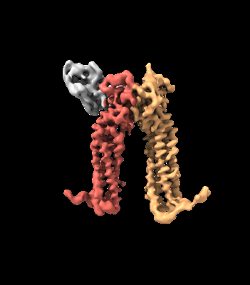 emd_14991.png emd_14991.png | 51.3 KB | ||
| Filedesc metadata | emd-14991.cif.gz | 6.3 KB | ||
| Others | emd_14991_additional_1.map.gzemd_14991_half_map_1.map.gzemd_14991_half_map_2.map.gz | 256.2 MB 285.4 MB 285.4 MB | ||
| Archive directory |  http://ftp.pdbj.org/pub/emdb/structures/EMD-14991ftp://ftp.pdbj.org/pub/emdb/structures/EMD-14991 http://ftp.pdbj.org/pub/emdb/structures/EMD-14991ftp://ftp.pdbj.org/pub/emdb/structures/EMD-14991 | HTTPS FTP |
-Related structure data
| Related structure data |  7zw1MC M: atomic model generated by this map C: citing same article ( |
|---|---|
| Similar structure data |


-Links
| EMDB pages | EMDB (EBI/PDBe) / EMDataResource |
|---|---|
| Related items in Molecule of the Month |

-Map
| File | Download / File: emd_14991.map.gz / Format: CCP4 / Size: 307.5 MB / Type: IMAGE STORED AS FLOATING POINT NUMBER (4 BYTES) | ||||||||||||||||||||||||||||||||||||
|---|---|---|---|---|---|---|---|---|---|---|---|---|---|---|---|---|---|---|---|---|---|---|---|---|---|---|---|---|---|---|---|---|---|---|---|---|---|
| Annotation | Non-sharpened map | ||||||||||||||||||||||||||||||||||||
| Projections & slices | Image control
Images are generated by Spider. | ||||||||||||||||||||||||||||||||||||
| Voxel size | X=Y=Z: 0.656 Å | ||||||||||||||||||||||||||||||||||||
| Density |
| ||||||||||||||||||||||||||||||||||||
| Symmetry | Space group: 1 | ||||||||||||||||||||||||||||||||||||
| Details | EMDB XML:
|
 Z (Sec.)
 Y (Row.)
 X (Col.)


-Supplemental data
-Additional map: Sharpened map
| File | emd_14991_additional_1.map | ||||||||||||
|---|---|---|---|---|---|---|---|---|---|---|---|---|---|
| Annotation | Sharpened map | ||||||||||||
| Projections & Slices |
| ||||||||||||
| Density Histograms |


-Half map: #2
| File | emd_14991_half_map_1.map | ||||||||||||
|---|---|---|---|---|---|---|---|---|---|---|---|---|---|
| Projections & Slices |
| ||||||||||||
| Density Histograms |


-Half map: #1
| File | emd_14991_half_map_2.map | ||||||||||||
|---|---|---|---|---|---|---|---|---|---|---|---|---|---|
| Projections & Slices |
| ||||||||||||
| Density Histograms |


- Sample components
-Entire : Human PRPH2-ROM1 hetero-dimer
| Entire | Name: Human PRPH2-ROM1 hetero-dimer |
|---|---|
| Components |
|
-Supramolecule #1: Human PRPH2-ROM1 hetero-dimer
| Supramolecule | Name: Human PRPH2-ROM1 hetero-dimer / type: complex / ID: 1 / Parent: 0 / Macromolecule list: all |
|---|---|
| Molecular weight | Theoretical: 76 kDa/nm |
-Supramolecule #2: Human PRPH2-ROM1 hetero-dimer
| Supramolecule | Name: Human PRPH2-ROM1 hetero-dimer / type: complex / ID: 2 / Parent: 1 / Macromolecule list: #1-#2 |
|---|---|
| Source (natural) | Organism: Homo sapiens (human) |
-Supramolecule #3: Nanobody
| Supramolecule | Name: Nanobody / type: complex / ID: 3 / Parent: 1 / Macromolecule list: #3 |
|---|---|
| Source (natural) | Organism: |
-Macromolecule #1: Peripherin-2
| Macromolecule | Name: Peripherin-2 / type: protein_or_peptide / ID: 1 Details: HHHHHH = tag of purification Sequence of the protein starts at A-L-L (Aminoacids 2-3-4) Number of copies: 1 / Enantiomer: LEVO |
|---|---|
| Source (natural) | Organism: Homo sapiens (human) |
| Molecular weight | Theoretical: 40.054137 KDa |
| Recombinant expression | Organism: Homo sapiens (human) |
| Sequence | String: HHHHHHGSAL LKVKFDQKKR VKLAQGLWLM NWFSVLAGII IFSLGLFLKI ELRKRSDVMN NSESHFVPNS LIGMGVLSCV FNSLAGKIC YDALDPAKYA RWKPWLKPYL AICVLFNIIL FLVALCCFLL RGSLENTLGQ GLKNGMKYYR DTDTPGRSFM K KTIDMLQI ...String: HHHHHHGSAL LKVKFDQKKR VKLAQGLWLM NWFSVLAGII IFSLGLFLKI ELRKRSDVMN NSESHFVPNS LIGMGVLSCV FNSLAGKIC YDALDPAKYA RWKPWLKPYL AICVLFNIIL FLVALCCFLL RGSLENTLGQ GLKNGMKYYR DTDTPGRSFM K KTIDMLQI EFKCCGNNGF RDWFEIQWIS NRYLDFSSKE VKDRIKSNVD GRYLVDGVPF SCCNPSSPRP CIQYQITNNS AH YSYDHQT EELNLWVRGC RAALLSYYSS LMNSMGVVTL LIWLFEVTIT IGLRYLQTSL DGVSNPEESE SESEGWLLEK SVP ETWKAF LESVKKLGKG NQVEAEGAGA GQAPEAG UniProtKB: Peripherin-2 |
-Macromolecule #2: Rod outer segment membrane protein 1
| Macromolecule | Name: Rod outer segment membrane protein 1 / type: protein_or_peptide / ID: 2 Details: The protein sequence starts at A-P-V-L, residues 2-3-4-5 Number of copies: 1 / Enantiomer: LEVO |
|---|---|
| Source (natural) | Organism: Homo sapiens (human) |
| Molecular weight | Theoretical: 37.249828 KDa |
| Recombinant expression | Organism: Homo sapiens (human) |
| Sequence | String: GSAPVLPLVL PLQPRIRLAQ GLWLLSWLLA LAGGVILLCS GHLLVQLRHL GTFLAPSCQF PVLPQAALAA GAVALGTGLV GVGASRASL NAALYPPWRG VLGPLLVAGT AGGGGLLVVG LGLALALPGS LDEALEEGLV TALAHYKDTE VPGHCQAKRL V DELQLRYH ...String: GSAPVLPLVL PLQPRIRLAQ GLWLLSWLLA LAGGVILLCS GHLLVQLRHL GTFLAPSCQF PVLPQAALAA GAVALGTGLV GVGASRASL NAALYPPWRG VLGPLLVAGT AGGGGLLVVG LGLALALPGS LDEALEEGLV TALAHYKDTE VPGHCQAKRL V DELQLRYH CCGRHGYKDW FGVQWVSSRY LDPGDRDVAD RIQSNVEGLY LTDGVPFSCC NPHSPRPCLQ NRLSDSYAHP LF DPRQPNQ NLWAQGCHEV LLEHLQDLAG TLGSMLAVTF LLQALVLLGL RYLQTALEGL GGVIDAGGET QGYLFPSGLK DML KTAWLQ GGVACRPAPE EAPPGEAPPK EDLSEA UniProtKB: Rod outer segment membrane protein 1 |
-Macromolecule #3: Nanobody
| Macromolecule | Name: Nanobody / type: protein_or_peptide / ID: 3 / Number of copies: 1 / Enantiomer: LEVO |
|---|---|
| Source (natural) | Organism: |
| Molecular weight | Theoretical: 14.646134 KDa |
| Recombinant expression | Organism:  |
| Sequence | String: SQVQLQESGG GLVQAGGSLR LSCAASTRTT SRYTVGWFCQ APGKEREFVA AVHWSGGSTW YADSVKGRFT ISRDNAKNTV YLQMNSLKQ EDTAVYYCAA AEPRRYSYYM RPDEYNYWGQ GTQVTVSSAA PLE |
-Experimental details
-Structure determination
| Method | cryo EM |
|---|---|
 Processing Processing | single particle reconstruction |
| Aggregation state | particle |
-Sample preparation
| Buffer | pH: 7.6 |
|---|---|
| Vitrification | Cryogen name: ETHANE |
- Electron microscopy
| Microscope | FEI TITAN KRIOS |
|---|---|
| Image recording | Film or detector model: GATAN K3 BIOQUANTUM (6k x 4k) / Average electron dose: 60.0 e/Å2 |
| Electron beam | Acceleration voltage: 300 kV / Electron source:  FIELD EMISSION GUN FIELD EMISSION GUN |
| Electron optics | Illumination mode: OTHER / Imaging mode: BRIGHT FIELD / Nominal defocus max: 2.3000000000000003 µm / Nominal defocus min: 0.8 µm |
| Experimental equipment |  Model: Titan Krios / Image courtesy: FEI Company |

