Organelle or cellular component: Secretory phospholipase A2 receptor
Protein or peptide: Secretory phospholipase A2 receptor
Keywords
Membranous nephropathy / IMMUNE SYSTEM
Function / homology
Function and homology information
negative regulation of arachidonate secretion / Acyl chain remodelling of PG / Acyl chain remodelling of PC / Acyl chain remodelling of PI / Acyl chain remodelling of PS / Acyl chain remodelling of PE / phospholipase A2 inhibitor activity / positive regulation of podocyte apoptotic process / Synthesis of PA / positive regulation of arachidonate secretion ...negative regulation of arachidonate secretion / Acyl chain remodelling of PG / Acyl chain remodelling of PC / Acyl chain remodelling of PI / Acyl chain remodelling of PS / Acyl chain remodelling of PE / phospholipase A2 inhibitor activity / positive regulation of podocyte apoptotic process / Synthesis of PA / positive regulation of arachidonate secretion / oxidative stress-induced premature senescence / positive regulation of DNA damage response, signal transduction by p53 class mediator / replicative senescence / phospholipase binding / reactive oxygen species metabolic process / receptor-mediated endocytosis / positive regulation of cytokine production / carbohydrate binding / signaling receptor activity / signaling receptor complex / cell surface / extracellular region / plasma membrane Similarity search - Function
MRC2-like, N-terminal cysteine-rich domain / Fibronectin type II domain / Fibronectin type II domain superfamily / Fibronectin type II domain / Fibronectin type-II collagen-binding domain signature. / Fibronectin type-II collagen-binding domain profile. / Fibronectin type 2 domain / : / C-type lectin, conserved site / C-type lectin domain signature. ...MRC2-like, N-terminal cysteine-rich domain / Fibronectin type II domain / Fibronectin type II domain superfamily / Fibronectin type II domain / Fibronectin type-II collagen-binding domain signature. / Fibronectin type-II collagen-binding domain profile. / Fibronectin type 2 domain / : / C-type lectin, conserved site / C-type lectin domain signature. / Ricin-type beta-trefoil / Lectin domain of ricin B chain profile. / Ricin B, lectin domain / Ricin B-like lectins / Lectin C-type domain / C-type lectin domain profile. / C-type lectin-like / C-type lectin (CTL) or carbohydrate-recognition domain (CRD) / C-type lectin-like/link domain superfamily / C-type lectin fold Similarity search - Domain/homology
Journal: Proc Natl Acad Sci U S A / Year: 2022 Title: Structure of PLA2R reveals presentation of the dominant membranous nephropathy epitope and an immunogenic patch. Authors: Maryline Fresquet / Michael P Lockhart-Cairns / Samuel J Rhoden / Thomas A Jowitt / David C Briggs / Clair Baldock / Paul E Brenchley / Rachel Lennon / Abstract: Membranous nephropathy is an autoimmune kidney disease caused by autoantibodies targeting antigens present on glomerular podocytes, instigating a cascade leading to glomerular injury. The most ...Membranous nephropathy is an autoimmune kidney disease caused by autoantibodies targeting antigens present on glomerular podocytes, instigating a cascade leading to glomerular injury. The most prevalent circulating autoantibodies in membranous nephropathy are against phospholipase A2 receptor (PLA2R), a cell surface receptor. The dominant epitope in PLA2R is located within the cysteine-rich domain, yet high-resolution structure-based mapping is lacking. In this study, we define the key nonredundant amino acids in the dominant epitope of PLA2R involved in autoantibody binding. We further describe two essential regions within the dominant epitope and spacer requirements for a synthetic peptide of the epitope for drug discovery. In addition, using cryo-electron microscopy, we have determined the high-resolution structure of PLA2R to 3.4 Å resolution, which shows that the dominant epitope and key residues within the cysteine-rich domain are accessible at the cell surface. In addition, the structure of PLA2R not only suggests a different orientation of domains but also implicates a unique immunogenic signature in PLA2R responsible for inducing autoantibody formation and recognition.
EMPIAR-10915 (Title: CryoEM data of PLA2R at pH 6.2 with both 0 and 30 degree tilts. Data size: 2.1 TB Data #1: Unaligned multi-frame images of PLA2R at pH 6.2 with a 30 degree tilt. [micrographs - multiframe] Data #2: Unaligned multi-frame images of PLA2R at pH 6.2 with 0 degree tilt (not used in publication) [micrographs - multiframe])
Model: Quantifoil R1.2/1.3 / Material: COPPER / Mesh: 300 / Support film - Material: CARBON / Support film - topology: HOLEY / Pretreatment - Type: GLOW DISCHARGE / Pretreatment - Time: 25 sec. / Pretreatment - Atmosphere: AIR
Vitrification
Cryogen name: ETHANE / Chamber humidity: 100 % / Chamber temperature: 277 K / Instrument: FEI VITROBOT MARK IV Details: Sample were applied to the grid in the chamber and blotted for 3 seconds..
Details
Sample was purified from secreted mammalian media with Ni affinity. The sample was further purified with SEC and buffer exchanged to pH 6.2.
-
Electron microscopy
Microscope
TFS KRIOS
Specialist optics
Energy filter - Name: GIF Bioquantum / Energy filter - Slit width: 20 eV
Details
30 degree tilt of stage
Image recording
Film or detector model: GATAN K3 (6k x 4k) / Number grids imaged: 1 / Average exposure time: 2.0 sec. / Average electron dose: 42.9 e/Å2
Electron beam
Acceleration voltage: 300 kV / Electron source: FIELD EMISSION GUN
In the structure databanks used in Yorodumi, some data are registered as the other names, "COVID-19 virus" and "2019-nCoV". Here are the details of the virus and the list of structure data.
Jan 31, 2019. EMDB accession codes are about to change! (news from PDBe EMDB page)
EMDB accession codes are about to change! (news from PDBe EMDB page)
The allocation of 4 digits for EMDB accession codes will soon come to an end. Whilst these codes will remain in use, new EMDB accession codes will include an additional digit and will expand incrementally as the available range of codes is exhausted. The current 4-digit format prefixed with “EMD-” (i.e. EMD-XXXX) will advance to a 5-digit format (i.e. EMD-XXXXX), and so on. It is currently estimated that the 4-digit codes will be depleted around Spring 2019, at which point the 5-digit format will come into force.
The EM Navigator/Yorodumi systems omit the EMD- prefix.
Related info.:Q: What is EMD? / ID/Accession-code notation in Yorodumi/EM Navigator
Yorodumi is a browser for structure data from EMDB, PDB, SASBDB, etc.
This page is also the successor to EM Navigator detail page, and also detail information page/front-end page for Omokage search.
The word "yorodu" (or yorozu) is an old Japanese word meaning "ten thousand". "mi" (miru) is to see.
Related info.:EMDB / PDB / SASBDB / Comparison of 3 databanks / Yorodumi Search / Aug 31, 2016. New EM Navigator & Yorodumi / Yorodumi Papers / Jmol/JSmol / Function and homology information / Changes in new EM Navigator and Yorodumi
 Movie
Movie Controller


 Open data
 Basic information
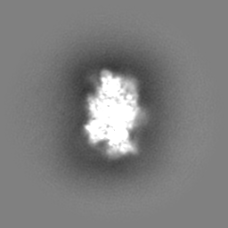
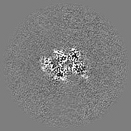
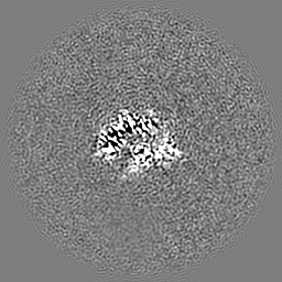
 Map data
 Sample
 Keywords
 Function and homology information
 Homo sapiens (human)
 Authors
 United Kingdom, 5 items
  Citation
 Structure visualization
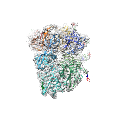
 Downloads & links
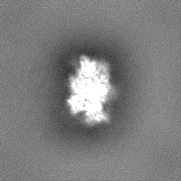
 emd_14077.png
 http://ftp.pdbj.org/pub/emdb/structures/EMD-14077


 Z (Sec.)
 Y (Row.)
 X (Col.)


 Sample components
 Processing
 Electron microscopy
 FIELD EMISSION GUN
