ジャーナル: Elife / 年: 2021 タイトル: Electron cryo-tomography reveals the subcellular architecture of growing axons in human brain organoids. 著者: Patrick C Hoffmann / Stefano L Giandomenico / Iva Ganeva / Michael R Wozny / Magdalena Sutcliffe / Madeline A Lancaster / Wanda Kukulski / 要旨: During brain development, axons must extend over great distances in a relatively short amount of time. How the subcellular architecture of the growing axon sustains the requirements for such rapid ...During brain development, axons must extend over great distances in a relatively short amount of time. How the subcellular architecture of the growing axon sustains the requirements for such rapid build-up of cellular constituents has remained elusive. Human axons have been particularly poorly accessible to imaging at high resolution in a near-native context. Here, we present a method that combines cryo-correlative light microscopy and electron tomography with human cerebral organoid technology to visualize growing axon tracts. Our data reveal a wealth of structural details on the arrangement of macromolecules, cytoskeletal components, and organelles in elongating axon shafts. In particular, the intricate shape of the endoplasmic reticulum is consistent with its role in fulfilling the high demand for lipid biosynthesis to support growth. Furthermore, the scarcity of ribosomes within the growing shaft suggests limited translational competence during expansion of this compartment. These findings establish our approach as a powerful resource for investigating the ultrastructure of defined neuronal compartments.
EMPIAR-10804 (タイトル: electron cryo-tomograms of axons from human cerebral organoids, expressing GFP-ESYT1 Data size: 40.9 Data #1: reconstructed electron tomographic volumes of axons from cerebral organoids expressing GFP-ESYT1 [reconstructed volumes] Data #2: Unaligned raw image frames of tilt series acquired on axons from cerebral organoids expressing GFP-ESYT1 [micrographs - multiframe])
 ムービー
ムービー コントローラー


 データを開く
 基本情報
基本情報 マップデータ
 試料
試料 Homo sapiens (ヒト)
 データ登録者
 英国, European Union, 3件
  引用
引用
 構造の表示
構造の表示 ムービービューア
 ダウンロードとリンク
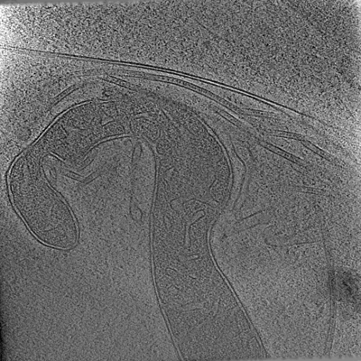 emd_13197.png
 http://ftp.pdbj.org/pub/emdb/structures/EMD-13197
 試料の構成要素
 解析
解析 電子顕微鏡法
 FIELD EMISSION GUN
