- EMDB-13056: Tomogram of a nuclease treated shVim HO H222P MEF nuclear lamina -
+
データを開く
IDまたはキーワード:
読み込み中...
-
基本情報
登録情報
データベース: EMDB / ID: EMD-13056
タイトル
Tomogram of a nuclease treated shVim HO H222P MEF nuclear lamina
マップデータ
A tomogram of nuclease treated shvim HO H222P MEF nucleus. The tomogram is dose-weighted, CTF-corrected and reconstructed in IMOD. Fudicial markers are replaced by noise.
ジャーナル: J Cell Sci / 年: 2021 タイトル: A lamin A/C variant causing striated muscle disease provides insights into filament organization. 著者: Rafael Kronenberg-Tenga / Meltem Tatli / Matthias Eibauer / Wei Wu / Ji-Yeon Shin / Gisèle Bonne / Howard J Worman / Ohad Medalia / 要旨: The gene encodes the A-type lamins, which polymerize into ∼3.5-nm-thick filaments and, together with B-type lamins and associated proteins, form the nuclear lamina. Mutations in cause a wide ...The gene encodes the A-type lamins, which polymerize into ∼3.5-nm-thick filaments and, together with B-type lamins and associated proteins, form the nuclear lamina. Mutations in cause a wide variety of pathologies. In this study, we analyzed the nuclear lamina of embryonic fibroblasts from mice, which develop cardiomyopathy and muscular dystrophy. Although the organization of the lamina appeared unaltered, there were changes in chromatin and B-type lamin expression. An increase in nuclear size and consequently a relative reduction in heterochromatin near the lamina allowed for a higher resolution structural analysis of lamin filaments using cryo-electron tomography. This was most apparent when visualizing lamin filaments and using a nuclear extraction protocol. Averaging of individual segments of filaments in mouse fibroblasts resolved two polymers that constitute the mature filaments. Our findings provide better views of the organization of lamin filaments and the effect of a striated muscle disease-causing mutation on nuclear structure.
EMPIAR-10601 (タイトル: A lamin A/C variant causing striated muscle disease provides insights into filament organization Data size: 2.7 Data #1: Tilt-series of LmnaH222P/H222P revealing lamin meshwork in nuclease treated and FIB-milled nucleus [tilt series])
ダウンロード / ファイル: emd_13056.map.gz / 形式: CCP4 / 大きさ: 165.7 MB / タイプ: IMAGE STORED AS SIGNED BYTE
注釈
A tomogram of nuclease treated shvim HO H222P MEF nucleus. The tomogram is dose-weighted, CTF-corrected and reconstructed in IMOD. Fudicial markers are replaced by noise.
ボクセルのサイズ
X=Y=Z: 8.82603 Å
密度
最小 - 最大
-128.0 - 127.0
平均 (標準偏差)
22.722765 (±17.823496)
対称性
空間群: 1
詳細
EMDB XML:
マップ形状
Axis order
X
Y
Z
Origin
16
-16
-98
サイズ
928
960
195
Spacing
960
928
195
セル
A: 8472.988 Å / B: 8190.5557 Å / C: 1721.0758 Å α=β=γ: 90.0 °
CCP4マップ ヘッダ情報:
mode
envelope stored as signed bytes (from -128 lowest to 127 highest)
 ムービー
ムービー コントローラー


 データを開く
 基本情報
基本情報 マップデータ
 試料
試料
 データ登録者
 スイス, 1件
  引用
引用

 構造の表示
構造の表示 ムービービューア
 ダウンロードとリンク
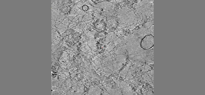 emd_13056.png
 http://ftp.pdbj.org/pub/emdb/structures/EMD-13056
 試料の構成要素
 解析
解析 電子顕微鏡法
 FIELD EMISSION GUN
