Movie
Movie Controller


+ Open data

- Basic information
| Entry | Database: EMDB / ID: EMD-10736 | |||||||||
|---|---|---|---|---|---|---|---|---|---|---|
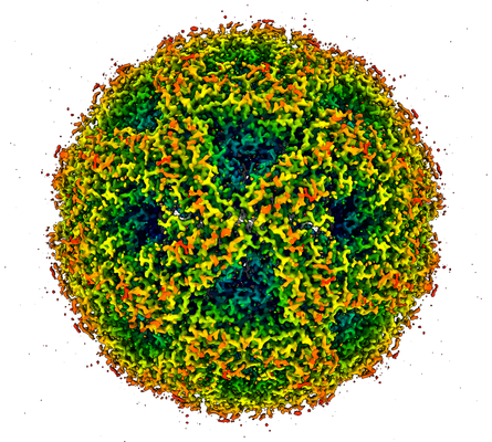
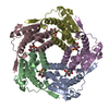
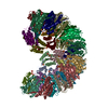
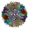
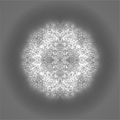
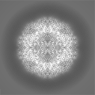
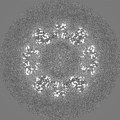
| Title | Lumazine Synthase | |||||||||
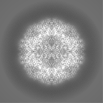
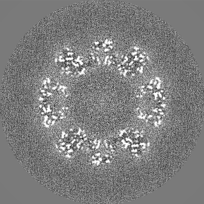
 Map data Map data | Reconstruction of Lumazine Synthase, sharpened and filtered at 2 angstroms | |||||||||
 Sample Sample |
| |||||||||
| Biological species |   Aquifex aeolicus (bacteria) Aquifex aeolicus (bacteria) | |||||||||
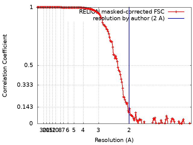
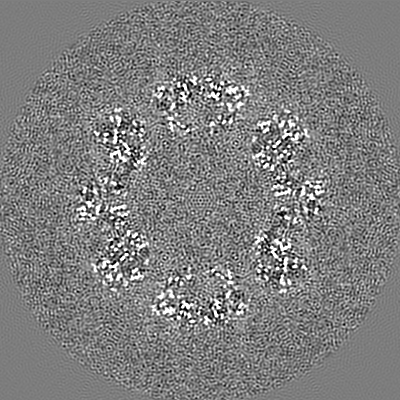
| Method | single particle reconstruction / cryo EM / Resolution: 2.0 Å | |||||||||
 Authors Authors | Bhella D / Streetley J / Clarke M / Cowton V / Patel A / Bammes B / Hosogi N | |||||||||
| Funding support |  United Kingdom, 2 items United Kingdom, 2 items
| |||||||||
 Citation Citation | Journal: Biophys Rev / Year: 2019 Title: Cryo-electron microscopy: an introduction to the technique, and considerations when working to establish a national facility. Authors: David Bhella / | |||||||||
| History |
|
- Structure visualization
| Movie |
 Movie viewer Movie viewer |
|---|---|
| Structure viewer | EM map: SurfViewMolmilJmol/JSmol |
| Supplemental images |
- Downloads & links
-EMDB archive
| Map data | emd_10736.map.gz | 227.1 MB | EMDB map data format | |
|---|---|---|---|---|
| Header (meta data) | emd-10736-v30.xmlemd-10736.xml | 16 KB 16 KB | Display Display | EMDB header |
| FSC (resolution estimation) | emd_10736_fsc.xml | 14.2 KB | Display | FSC data file |
| Images |  emd_10736.png emd_10736.png | 251.7 KB | ||
| Masks | emd_10736_msk_1.map | 244.1 MB | Mask map | |
| Others | emd_10736_half_map_1.map.gzemd_10736_half_map_2.map.gz | 193.5 MB 193.5 MB | ||
| Archive directory |  http://ftp.pdbj.org/pub/emdb/structures/EMD-10736ftp://ftp.pdbj.org/pub/emdb/structures/EMD-10736 http://ftp.pdbj.org/pub/emdb/structures/EMD-10736ftp://ftp.pdbj.org/pub/emdb/structures/EMD-10736 | HTTPS FTP |
-Related structure data
| Similar structure data | |
|---|---|
| EM raw data | EMPIAR-10384 (Title: Cryo-EM structure of Lumazine Synthase / Data size: 3.0 TB Data #1: Frame aligned multi-frame movies of Lumazine Synthase imaged on a CryoARM300 and DE64 [micrographs - multiframe]) EMPIAR-10385 (Title: Cryo-EM structure of Lumazine Synthase / Data size: 6.8 TBData #1: Frame aligned multi-frame movies of Lumazine Synthase imaged on a CryoARM300 and DE64 [micrographs - multiframe]) |

-Links
| EMDB pages | EMDB (EBI/PDBe) / EMDataResource |
|---|
-Map
| File | Download / File: emd_10736.map.gz / Format: CCP4 / Size: 244.1 MB / Type: IMAGE STORED AS FLOATING POINT NUMBER (4 BYTES) | ||||||||||||||||||||||||||||||||||||||||||||||||||||||||||||
|---|---|---|---|---|---|---|---|---|---|---|---|---|---|---|---|---|---|---|---|---|---|---|---|---|---|---|---|---|---|---|---|---|---|---|---|---|---|---|---|---|---|---|---|---|---|---|---|---|---|---|---|---|---|---|---|---|---|---|---|---|---|
| Annotation | Reconstruction of Lumazine Synthase, sharpened and filtered at 2 angstroms | ||||||||||||||||||||||||||||||||||||||||||||||||||||||||||||
| Projections & slices | Image control
Images are generated by Spider. | ||||||||||||||||||||||||||||||||||||||||||||||||||||||||||||
| Voxel size | X=Y=Z: 0.597 Å | ||||||||||||||||||||||||||||||||||||||||||||||||||||||||||||
| Density |
| ||||||||||||||||||||||||||||||||||||||||||||||||||||||||||||
| Symmetry | Space group: 1 | ||||||||||||||||||||||||||||||||||||||||||||||||||||||||||||
| Details | EMDB XML:
CCP4 map header:
| ||||||||||||||||||||||||||||||||||||||||||||||||||||||||||||
 Z (Sec.)
 Y (Row.)
 X (Col.)


-Supplemental data
-Mask #1
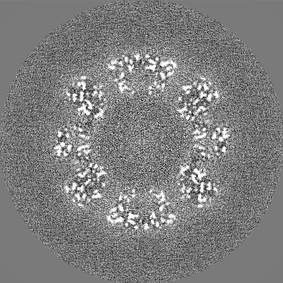
| File | emd_10736_msk_1.map | ||||||||||||
|---|---|---|---|---|---|---|---|---|---|---|---|---|---|
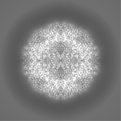
| Projections & Slices |
| ||||||||||||
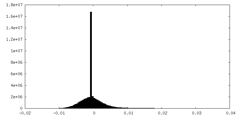
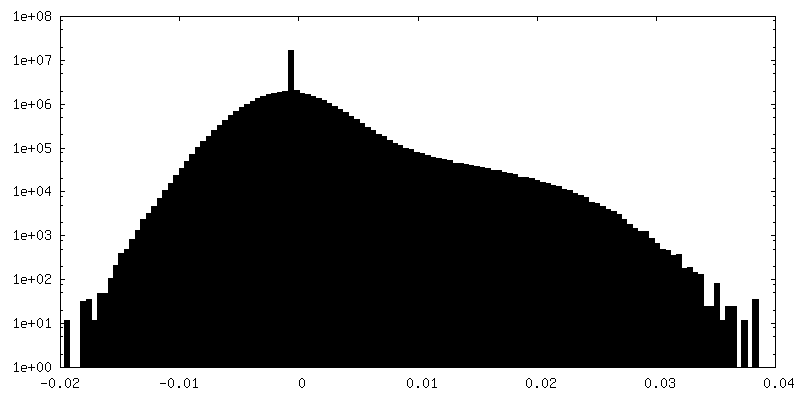
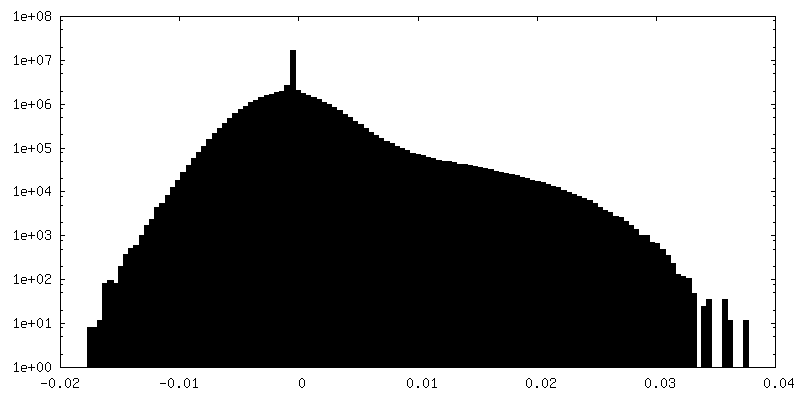
| Density Histograms |

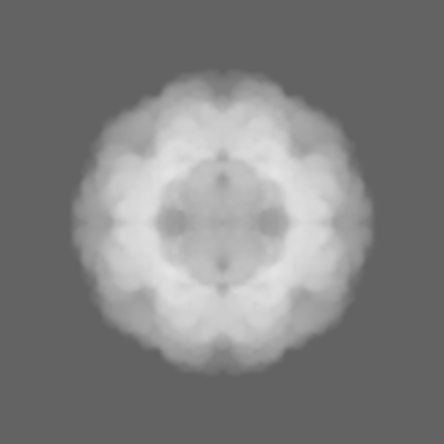


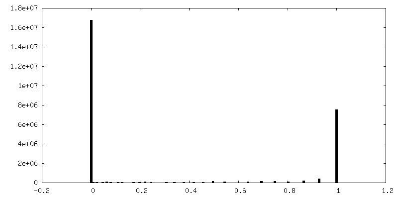
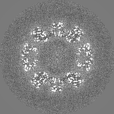
-Half map: Half map 1
| File | emd_10736_half_map_1.map | ||||||||||||
|---|---|---|---|---|---|---|---|---|---|---|---|---|---|
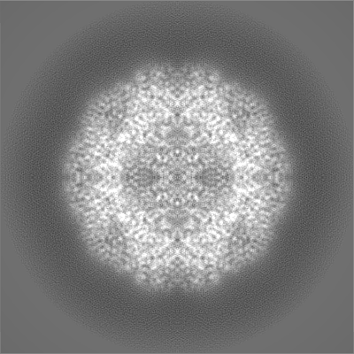
| Annotation | Half map 1 | ||||||||||||
| Projections & Slices |
| ||||||||||||
| Density Histograms |

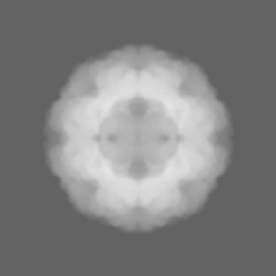
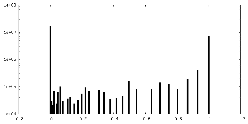
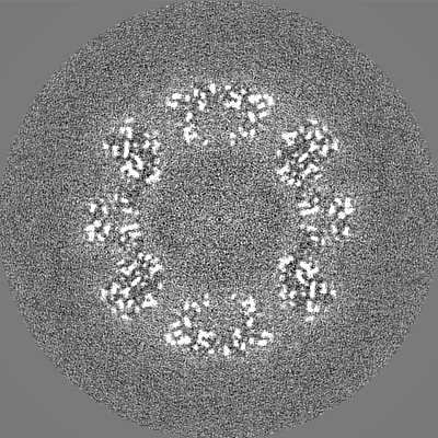
-Half map: Half map 2
| File | emd_10736_half_map_2.map | ||||||||||||
|---|---|---|---|---|---|---|---|---|---|---|---|---|---|
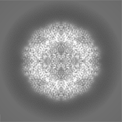
| Annotation | Half map 2 | ||||||||||||
| Projections & Slices |
| ||||||||||||
| Density Histograms |

- Sample components
-Entire : Icosahedral complex of Lumazine Synthase
| Entire | Name: Icosahedral complex of Lumazine Synthase |
|---|---|
| Components |
|
-Supramolecule #1: Icosahedral complex of Lumazine Synthase
| Supramolecule | Name: Icosahedral complex of Lumazine Synthase / type: complex / ID: 1 / Parent: 0 |
|---|---|
| Source (natural) | Organism: Aquifex aeolicus (bacteria) |
| Recombinant expression | Organism: |
-Experimental details
-Structure determination
| Method | cryo EM |
|---|---|
 Processing Processing | single particle reconstruction |
| Aggregation state | particle |
-Sample preparation
| Concentration | 3 mg/mL |
|---|---|
| Buffer | pH: 7.2 |
| Grid | Model: C-flat-1.2/1.3 4C / Material: COPPER / Mesh: 400 / Support film - Material: CARBON / Support film - topology: HOLEY ARRAY / Pretreatment - Type: GLOW DISCHARGE |
| Vitrification | Cryogen name: ETHANE / Instrument: FEI VITROBOT MARK IV |
| Details | Purified by immobilised metal affinity chromatography. |
- Electron microscopy
| Microscope | JEOL CRYO ARM 300 |
|---|---|
| Specialist optics | Energy filter - Name: In-column Omega Filter / Energy filter - Slit width: 20 eV |
| Image recording | Film or detector model: DIRECT ELECTRON DE-64 (8k x 8k) / Detector mode: COUNTING / Digitization - Dimensions - Width: 4096 pixel / Digitization - Dimensions - Height: 4096 pixel / Digitization - Sampling interval: 13.0 µm / Digitization - Frames/image: 1-41 / Number grids imaged: 1 / Number real images: 1199 / Average exposure time: 10.0 sec. / Average electron dose: 1.66 e/Å2 Details: Images were acquired at 141 FPS with 2 x binning and electron counting. A total of 41 summed frames were written out. |
| Electron beam | Acceleration voltage: 300 kV / Electron source:  FIELD EMISSION GUN FIELD EMISSION GUN |
| Electron optics | C2 aperture diameter: 100.0 µm / Illumination mode: FLOOD BEAM / Imaging mode: BRIGHT FIELD / Cs: 2.7 mm / Nominal magnification: 200000 |
| Sample stage | Specimen holder model: JEOL CRYOSPECPORTER / Cooling holder cryogen: NITROGEN |