 Movie
Movie Controller


+ Open data

- Basic information
| Entry | Database: EMDB / ID: EMD-10160 | |||||||||
|---|---|---|---|---|---|---|---|---|---|---|
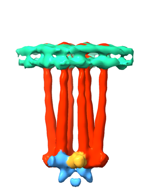
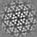
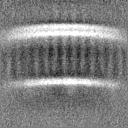
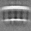
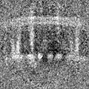
| Title | In Situ Core-Signalling Unit of E. coli Chemoreceptor Array | |||||||||
 Map data Map data | None | |||||||||
 Sample Sample |
| |||||||||
| Biological species |  | |||||||||
| Method | subtomogram averaging / cryo EM / Resolution: 16.0 Å | |||||||||
 Authors Authors | Burt A / Desfosses A / Gutsche I | |||||||||
| Funding support | 1 items
| |||||||||
 Citation Citation | Journal: Nat Commun / Year: 2020 Title: Complete structure of the chemosensory array core signalling unit in an E. coli minicell strain. Authors: Alister Burt / C Keith Cassidy / Peter Ames / Maria Bacia-Verloop / Megghane Baulard / Karine Huard / Zaida Luthey-Schulten / Ambroise Desfosses / Phillip J Stansfeld / William Margolin / ...Authors: Alister Burt / C Keith Cassidy / Peter Ames / Maria Bacia-Verloop / Megghane Baulard / Karine Huard / Zaida Luthey-Schulten / Ambroise Desfosses / Phillip J Stansfeld / William Margolin / John S Parkinson / Irina Gutsche /    Abstract: Motile bacteria sense chemical gradients with transmembrane receptors organised in supramolecular signalling arrays. Understanding stimulus detection and transmission at the molecular level requires ...Motile bacteria sense chemical gradients with transmembrane receptors organised in supramolecular signalling arrays. Understanding stimulus detection and transmission at the molecular level requires precise structural characterisation of the array building block known as a core signalling unit. Here we introduce an Escherichia coli strain that forms small minicells possessing extended and highly ordered chemosensory arrays. We use cryo-electron tomography and subtomogram averaging to provide a three-dimensional map of a complete core signalling unit, with visible densities corresponding to the HAMP and periplasmic domains. This map, combined with previously determined high resolution structures and molecular dynamics simulations, yields a molecular model of the transmembrane core signalling unit and enables spatial localisation of its individual domains. Our work thus offers a solid structural basis for the interpretation of a wide range of existing data and the design of further experiments to elucidate signalling mechanisms within the core signalling unit and larger array. | |||||||||
| History |
|
- Structure visualization
| Movie |
 Movie viewer Movie viewer |
|---|---|
| Structure viewer | EM map: SurfViewMolmilJmol/JSmol |
| Supplemental images |
- Downloads & links
-EMDB archive
| Map data | emd_10160.map.gz | 4.8 MB | EMDB map data format | |
|---|---|---|---|---|
| Header (meta data) | emd-10160-v30.xmlemd-10160.xml | 16.9 KB 16.9 KB | Display Display | EMDB header |
| FSC (resolution estimation) | emd_10160_fsc.xml | 4.7 KB | Display | FSC data file |
| Images |  emd_10160.png emd_10160.png | 55.5 KB | ||
| Masks | emd_10160_msk_1.mapemd_10160_msk_2.map | 8 MB 8 MB | Mask map | |
| Others | emd_10160_additional.map.gzemd_10160_additional_1.map.gzemd_10160_half_map_1.map.gzemd_10160_half_map_2.map.gz | 4.5 MB 4.5 MB 7.4 MB 7.4 MB | ||
| Archive directory |  http://ftp.pdbj.org/pub/emdb/structures/EMD-10160ftp://ftp.pdbj.org/pub/emdb/structures/EMD-10160 http://ftp.pdbj.org/pub/emdb/structures/EMD-10160ftp://ftp.pdbj.org/pub/emdb/structures/EMD-10160 | HTTPS FTP |
-Related structure data
| Similar structure data | |
|---|---|
| EM raw data | EMPIAR-10364 (Title: Cryo-electron tomography of E. coli minicells / Data size: 141.7 Data #1: Unaligned multi-frame micrographs from tilt series containing WM4196 minicells [micrographs - multiframe]) |
-Links
| EMDB pages | EMDB (EBI/PDBe) / EMDataResource |
|---|
-Map
| File | Download / File: emd_10160.map.gz / Format: CCP4 / Size: 8 MB / Type: IMAGE STORED AS FLOATING POINT NUMBER (4 BYTES) | ||||||||||||||||||||||||||||||||||||||||||||||||||||||||||||
|---|---|---|---|---|---|---|---|---|---|---|---|---|---|---|---|---|---|---|---|---|---|---|---|---|---|---|---|---|---|---|---|---|---|---|---|---|---|---|---|---|---|---|---|---|---|---|---|---|---|---|---|---|---|---|---|---|---|---|---|---|---|
| Annotation | None | ||||||||||||||||||||||||||||||||||||||||||||||||||||||||||||
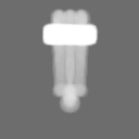
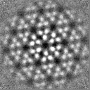
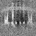
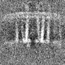
| Projections & slices | Image control
Images are generated by Spider. | ||||||||||||||||||||||||||||||||||||||||||||||||||||||||||||
| Voxel size | X=Y=Z: 4.48 Å | ||||||||||||||||||||||||||||||||||||||||||||||||||||||||||||
| Density |
| ||||||||||||||||||||||||||||||||||||||||||||||||||||||||||||
| Symmetry | Space group: 1 | ||||||||||||||||||||||||||||||||||||||||||||||||||||||||||||
| Details | EMDB XML:
CCP4 map header:
| ||||||||||||||||||||||||||||||||||||||||||||||||||||||||||||
 Z (Sec.)
 Y (Row.)
 X (Col.)


-Supplemental data
-Mask #1
| File | emd_10160_msk_1.map | ||||||||||||
|---|---|---|---|---|---|---|---|---|---|---|---|---|---|
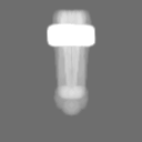
| Projections & Slices |
| ||||||||||||
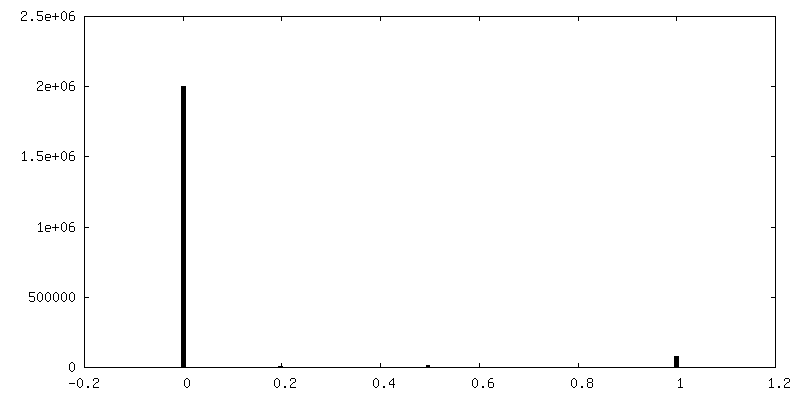
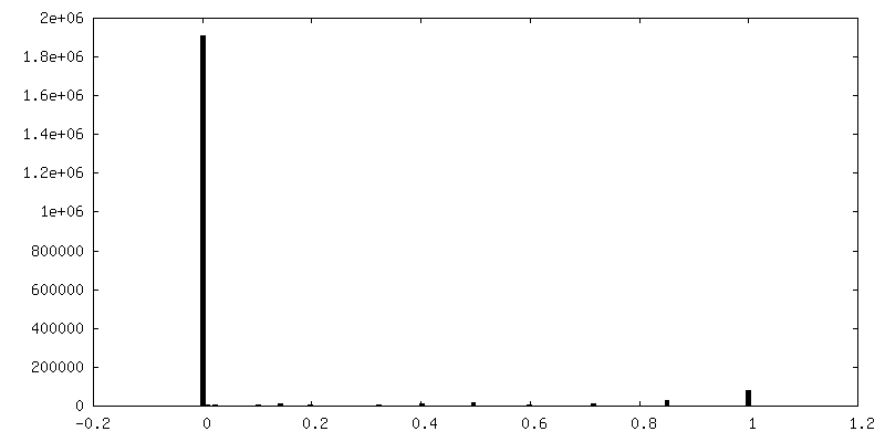
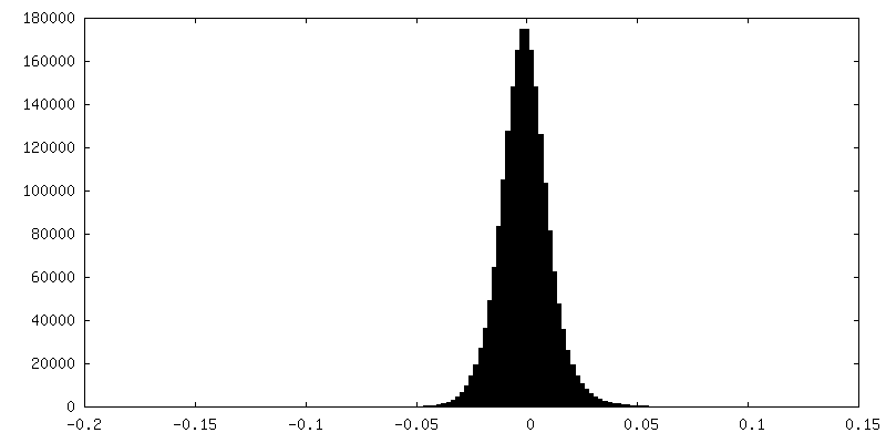
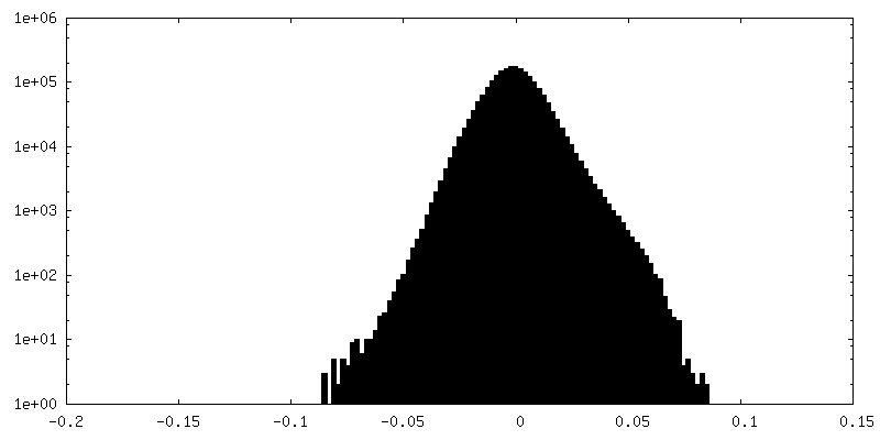
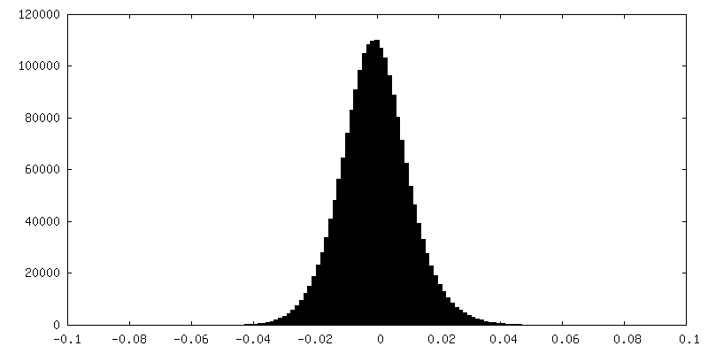
| Density Histograms |


-Mask #2
| File | emd_10160_msk_2.map | ||||||||||||
|---|---|---|---|---|---|---|---|---|---|---|---|---|---|
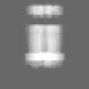
| Projections & Slices |
| ||||||||||||
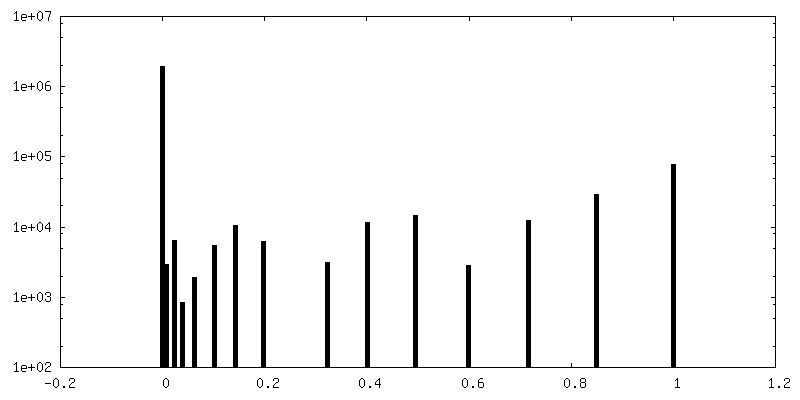
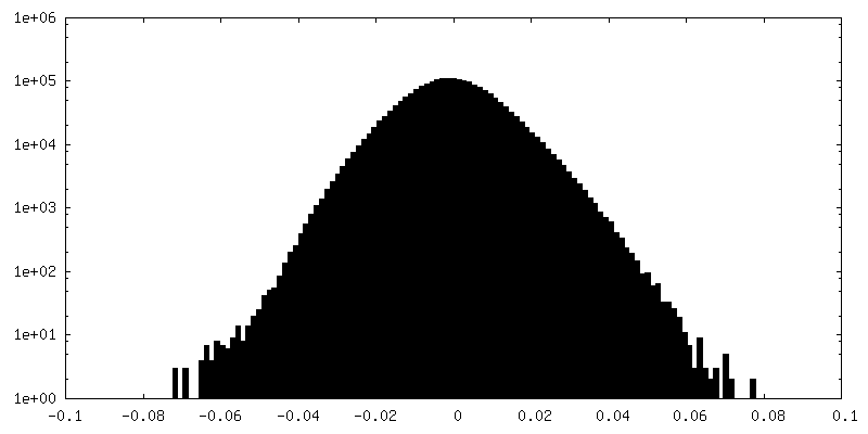
| Density Histograms |


-Additional map: Local resolution filtered map centered on 3fold axis...
| File | emd_10160_additional.map | ||||||||||||
|---|---|---|---|---|---|---|---|---|---|---|---|---|---|
| Annotation | Local resolution filtered map centered on 3fold axis of chemoreceptor array. | ||||||||||||
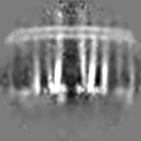
| Projections & Slices |
| ||||||||||||
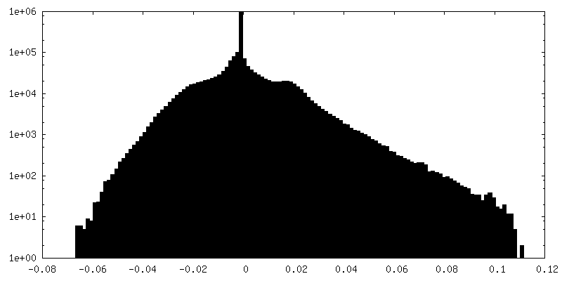
| Density Histograms |


-Additional map: Local resolution filtered map centered on 3fold axis...
| File | emd_10160_additional_1.map | ||||||||||||
|---|---|---|---|---|---|---|---|---|---|---|---|---|---|
| Annotation | Local resolution filtered map centered on 3fold axis of chemoreceptor array. | ||||||||||||
| Projections & Slices |
| ||||||||||||
| Density Histograms |


-Half map: even half map.
| File | emd_10160_half_map_1.map | ||||||||||||
|---|---|---|---|---|---|---|---|---|---|---|---|---|---|
| Annotation | even half map. | ||||||||||||
| Projections & Slices |
| ||||||||||||
| Density Histograms |


-Half map: odd half map.
| File | emd_10160_half_map_2.map | ||||||||||||
|---|---|---|---|---|---|---|---|---|---|---|---|---|---|
| Annotation | odd half map. | ||||||||||||
| Projections & Slices |
| ||||||||||||
| Density Histograms |

- Sample components
-Entire : Optimised E. coli minicell strain WM4196 containing native-state ...
| Entire | Name: Optimised E. coli minicell strain WM4196 containing native-state chemoreceptor arrays. |
|---|---|
| Components |
|
-Supramolecule #1: Optimised E. coli minicell strain WM4196 containing native-state ...
| Supramolecule | Name: Optimised E. coli minicell strain WM4196 containing native-state chemoreceptor arrays. type: cell / ID: 1 / Parent: 0 Details: Minicells isolated from WM4196 cell culture by differential centrifugation. |
|---|---|
| Source (natural) | Organism: |
-Experimental details
-Structure determination
| Method | cryo EM |
|---|---|
 Processing Processing | subtomogram averaging |
| Aggregation state | cell |
-Sample preparation
| Buffer | pH: 7 |
|---|---|
| Grid | Model: Quantifoil R3.5/1 / Material: COPPER/RHODIUM / Mesh: 300 / Support film - Material: CARBON / Support film - topology: HOLEY / Pretreatment - Type: GLOW DISCHARGE / Pretreatment - Atmosphere: AIR / Details: 25 mA |
| Vitrification | Cryogen name: ETHANE / Instrument: FEI VITROBOT MARK IV |
- Electron microscopy
| Microscope | FEI TITAN KRIOS |
|---|---|
| Specialist optics | Phase plate: VOLTA PHASE PLATE |
| Image recording | Film or detector model: GATAN K2 SUMMIT (4k x 4k) / Detector mode: COUNTING / Digitization - Frames/image: 1-5 / Number grids imaged: 1 / Average electron dose: 1.0 e/Å2 Details: Images were collected in movie mode, 5 frames per movie. |
| Electron beam | Acceleration voltage: 300 kV / Electron source:  FIELD EMISSION GUN FIELD EMISSION GUN |
| Electron optics | Illumination mode: FLOOD BEAM / Imaging mode: BRIGHT FIELD / Nominal defocus min: 0.0002 µm |
| Sample stage | Specimen holder model: FEI TITAN KRIOS AUTOGRID HOLDER / Cooling holder cryogen: NITROGEN |
| Experimental equipment |  Model: Titan Krios / Image courtesy: FEI Company |
-Image processing
| Final reconstruction | Applied symmetry - Point group: C2 (2 fold cyclic) / Algorithm: BACK PROJECTION / Resolution.type: BY AUTHOR / Resolution: 16.0 Å / Resolution method: FSC 0.143 CUT-OFF / Software - Name: Dynamo Details: C3 symmetry applied during refinement. Masks used containing varying extents of the array and the inner membrane were used. Number subtomograms used: 618 |
|---|---|
| Extraction | Number tomograms: 4 / Number images used: 618 / Software - Name: Dynamo |
| Final angle assignment | Type: NOT APPLICABLE / Software - Name: Dynamo |
| FSC plot (resolution estimation) | 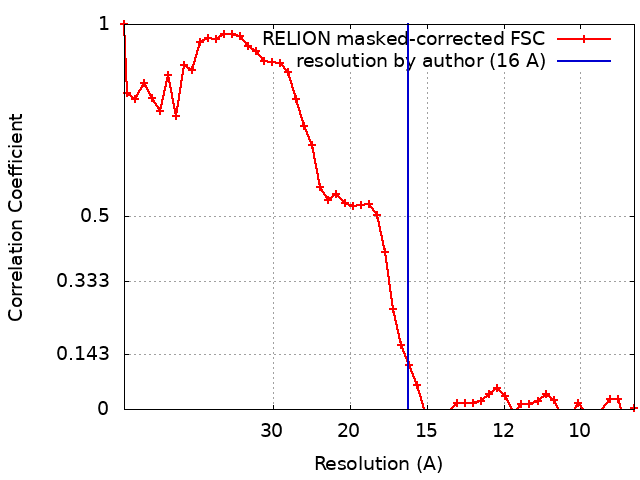 |
