 Movie
Movie Controller

[English] 日本語
 Yorodumi
- EMDB-0698: 120kV MicroED structure of FUS (37-42) SYSGYS solved from merged ... -
+ Open data

- Basic information
| Entry | Database: EMDB / ID: EMD-0698 | |||||||||||||||
|---|---|---|---|---|---|---|---|---|---|---|---|---|---|---|---|---|
| Title | 120kV MicroED structure of FUS (37-42) SYSGYS solved from merged datasets at 0.60 A | |||||||||||||||
 Map data Map data | 2Fo-Fc map | |||||||||||||||
 Sample Sample |
| |||||||||||||||
 Keywords Keywords | FUS / MicroED / Ultrahigh resolution / RNA BINDING PROTEIN | |||||||||||||||
| Function / homology |  Function and homology information Function and homology informationmembraneless organelle assembly / mRNA stabilization / mRNA Polyadenylation / regulation of RNA splicing / Dengue Virus-Host Interactions / Processing of Capped Intron-Containing Pre-mRNA / postsynaptic cytosol / positive regulation of double-strand break repair via homologous recombination / presynaptic cytosol / mRNA Splicing - Major Pathway ...membraneless organelle assembly / mRNA stabilization / mRNA Polyadenylation / regulation of RNA splicing / Dengue Virus-Host Interactions / Processing of Capped Intron-Containing Pre-mRNA / postsynaptic cytosol / positive regulation of double-strand break repair via homologous recombination / presynaptic cytosol / mRNA Splicing - Major Pathway / RNA splicing / transcription coregulator activity / mRNA 3'-UTR binding / molecular condensate scaffold activity / protein homooligomerization / GABA-ergic synapse / amyloid fibril formation / transcription coactivator activity / chromatin binding / regulation of transcription by RNA polymerase II / regulation of DNA-templated transcription / glutamatergic synapse / DNA binding / RNA binding / zinc ion binding / nucleoplasm / identical protein binding / nucleus Similarity search - Function | |||||||||||||||
| Biological species |  Homo sapiens (human) Homo sapiens (human) | |||||||||||||||
| Method | electron crystallography / cryo EM / Resolution: 0.6 Å | |||||||||||||||
 Authors Authors | Zhou H / Luo F | |||||||||||||||
| Funding support |  China, 4 items China, 4 items
| |||||||||||||||
 Citation Citation | Journal: Anal Chem / Year: 2019 Title: Programming Conventional Electron Microscopes for Solving Ultrahigh-Resolution Structures of Small and Macro-Molecules. Authors: Heng Zhou / Feng Luo / Zhipu Luo / Dan Li / Cong Liu / Xueming Li / Abstract: Microcrystal electron diffraction (MicroED) is becoming a powerful tool in determining the crystal structures of biological macromolecules and small organic compounds. However, wide applications of ...Microcrystal electron diffraction (MicroED) is becoming a powerful tool in determining the crystal structures of biological macromolecules and small organic compounds. However, wide applications of this technique are still limited by the special requirement for radiation-tolerated movie-mode camera and the lack of automated data collection methods. Herein, we develop a stage-camera synchronization scheme to minimize the hardware requirements and enable the use of the conventional electron cryo-microscope with a single-frame CCD camera, which ensures not only the acquisition of ultrahigh-resolution diffraction data but also low cost in practice. This method renders the structure determination of both peptide and small organic compounds at ultrahigh resolution up to ∼0.60 Å with unambiguous assignment of nearly all hydrogen atoms. The present work provides a widely applicable solution for routine structure determination of MicroED and demonstrates the capability of the low-end 120 kV microscope with a CCD camera in solving ultrahigh resolution structures of both organic compounds and biological macromolecules. | |||||||||||||||
| History |
|
- Structure visualization
| Movie |
Movie viewer |
|---|---|
| Structure viewer | EM map: SurfViewMolmilJmol/JSmol |
| Supplemental images |
 UCSF Chimera
- Downloads & links
-EMDB archive
| Map data | emd_0698.map.gz | 1.9 MB | EMDB map data format | |
|---|---|---|---|---|
| Header (meta data) | emd-0698-v30.xmlemd-0698.xml | 12.6 KB 12.6 KB | Display Display | EMDB header |
| Images |  emd_0698.png emd_0698.png | 47 KB | ||
| Filedesc metadata | emd-0698.cif.gz | 4.2 KB | ||
| Others | emd_0698_additional.map.gzemd_0698_additional_1.map.gz | 1.9 MB 1.9 MB | ||
| Filedesc structureFactors | emd_0698_sf.cif.gz | 133.9 KB | ||
| Archive directory |  http://ftp.pdbj.org/pub/emdb/structures/EMD-0698ftp://ftp.pdbj.org/pub/emdb/structures/EMD-0698 http://ftp.pdbj.org/pub/emdb/structures/EMD-0698ftp://ftp.pdbj.org/pub/emdb/structures/EMD-0698 | HTTPS FTP |
-Related structure data
| Related structure data |  6kj3MC 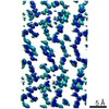 0696C  0697C 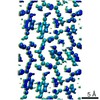 0699C 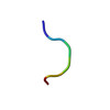 6kj1C  6kj2C  6kj4C M: atomic model generated by this map C: citing same article ( |
|---|---|
| Similar structure data | |
| EM raw data | EMPIAR-10319 (Title: 120kV MicroED structure of FUS (37-42) SYSGYS solved from merged datasets at 0.60 A Data size: 9.2 Data #1: 120kV MicroED data of FUS (37-42) SYSGYS [diffraction images]) |
-Links
| EMDB pages | EMDB (EBI/PDBe) / EMDataResource |
|---|---|
| Related items in Molecule of the Month |


-Map
| File | Download / File: emd_0698.map.gz / Format: CCP4 / Size: 10.9 MB / Type: IMAGE STORED AS FLOATING POINT NUMBER (4 BYTES) | ||||||||||||||||||||||||||||||||||||||||||||||||||||||||||||||||||||
|---|---|---|---|---|---|---|---|---|---|---|---|---|---|---|---|---|---|---|---|---|---|---|---|---|---|---|---|---|---|---|---|---|---|---|---|---|---|---|---|---|---|---|---|---|---|---|---|---|---|---|---|---|---|---|---|---|---|---|---|---|---|---|---|---|---|---|---|---|---|
| Annotation | 2Fo-Fc map | ||||||||||||||||||||||||||||||||||||||||||||||||||||||||||||||||||||
| Projections & slices | Image control
Images are generated by Spider. generated in cubic-lattice coordinate | ||||||||||||||||||||||||||||||||||||||||||||||||||||||||||||||||||||
| Voxel size | X: 0.1425 Å / Y: 0.14594 Å / Z: 0.14658 Å | ||||||||||||||||||||||||||||||||||||||||||||||||||||||||||||||||||||
| Density |
| ||||||||||||||||||||||||||||||||||||||||||||||||||||||||||||||||||||
| Symmetry | Space group: 1 | ||||||||||||||||||||||||||||||||||||||||||||||||||||||||||||||||||||
| Details | EMDB XML:
CCP4 map header:
| ||||||||||||||||||||||||||||||||||||||||||||||||||||||||||||||||||||
 X (Sec.)
 Y (Row.)
 Z (Col.)


-Supplemental data
-Additional map: Fo-Fc map
| File | emd_0698_additional.map | ||||||||||||
|---|---|---|---|---|---|---|---|---|---|---|---|---|---|
| Annotation | Fo-Fc map | ||||||||||||
| Projections & Slices |
| ||||||||||||
| Density Histograms |


-Additional map: Fo-Fc map
| File | emd_0698_additional_1.map | ||||||||||||
|---|---|---|---|---|---|---|---|---|---|---|---|---|---|
| Annotation | Fo-Fc map | ||||||||||||
| Projections & Slices |
| ||||||||||||
| Density Histograms |


- Sample components
-Entire : FUS LC RAC1
| Entire | Name: FUS LC RAC1 |
|---|---|
| Components |
|
-Supramolecule #1: FUS LC RAC1
| Supramolecule | Name: FUS LC RAC1 / type: complex / ID: 1 / Parent: 0 / Macromolecule list: #1 |
|---|
-Macromolecule #1: RNA-binding protein FUS
| Macromolecule | Name: RNA-binding protein FUS / type: protein_or_peptide / ID: 1 / Number of copies: 1 / Enantiomer: LEVO |
|---|---|
| Source (natural) | Organism: Homo sapiens (human) |
| Molecular weight | Theoretical: 662.648 Da |
| Sequence | String: SYSGYS UniProtKB: RNA-binding protein FUS |
-Macromolecule #2: water
| Macromolecule | Name: water / type: ligand / ID: 2 / Number of copies: 1 / Formula: HOH |
|---|---|
| Molecular weight | Theoretical: 18.015 Da |
| Chemical component information |  ChemComp-HOH: |
-Experimental details
-Structure determination
| Method | cryo EM |
|---|---|
 Processing Processing | electron crystallography |
| Aggregation state | 3D array |
-Sample preparation
| Buffer | pH: 7 |
|---|---|
| Vitrification | Cryogen name: ETHANE |
- Electron microscopy
| Microscope | FEI TECNAI 12 |
|---|---|
| Image recording | Film or detector model: FEI EAGLE (4k x 4k) / Average electron dose: 0.05 e/Å2 |
| Electron beam | Acceleration voltage: 120 kV / Electron source: LAB6 |
| Electron optics | Illumination mode: FLOOD BEAM / Imaging mode: DIFFRACTION / Camera length: 500 mm |
-Image processing
| Final reconstruction | Resolution.type: BY AUTHOR / Resolution: 0.6 Å / Resolution method: DIFFRACTION PATTERN/LAYERLINES |
|---|---|
| Crystallography statistics | Number intensities measured: 46057 / Number structure factors: 5850 / Fourier space coverage: 78.41 / R sym: 0.278 / R merge: 0.278 / Overall phase error: 44.58 / Overall phase residual: 1 / Phase error rejection criteria: 60 / High resolution: 0.6 Å / Shell - Shell ID: 1 / Shell - High resolution: 0.6 Å / Shell - Low resolution: 0.62 Å / Shell - Number structure factors: 474 / Shell - Phase residual: 1 / Shell - Fourier space coverage: 66.76 / Shell - Multiplicity: 3.94 |
-Atomic model buiding 1
| Refinement | Space: RECIPROCAL / Protocol: AB INITIO MODEL |
|---|---|
| Output model | PDB-6kj3: |
