 Movie
Movie Controller


+ Open data

- Basic information
| Entry | 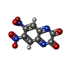 Database: PDB chemical components / ID: DNQ Database: PDB chemical components / ID: DNQ |
|---|---|
| Name | Name: |
-Chemical information
| Composition |  | ||||||
|---|---|---|---|---|---|---|---|
| Others | Type: NON-POLYMER / PDB classification: HETAIN / Three letter code: DNQ / Model coordinates PDB-ID: 1FTL | ||||||
| History |
| ||||||
 External links External links | UniChem / ChemSpider / BindingDB / CompTox / PubChem / SureChEMBL / Wikipedia search / Google search |
- Structure visualization
| Structure viewer | Molecule:  MolmilJmol/JSmol MolmilJmol/JSmol |
|---|
-Details
-SMILES
| ACDLabs 10.04 | [| CACTVS 3.341 | [ | OpenEye OEToolkits 1.5.0 | |
|---|
-SMILES CANONICAL
| CACTVS 3.341 | [| OpenEye OEToolkits 1.5.0 | |
|---|
-InChI
| InChI 1.03 |
|---|
-InChIKey
| InChI 1.03 |
|---|
-SYSTEMATIC NAME
| ACDLabs 10.04 | | OpenEye OEToolkits 1.5.0 | |
|---|
-PDB entries
Showing all 5 items

PDB-1ftl: 
CRYSTAL STRUCTURE OF THE GLUR2 LIGAND BINDING CORE (S1S2J) IN COMPLEX WITH THE ANTAGONIST DNQX AT 1.8 A RESOLUTION
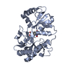
PDB-1lb9:
Crystal structure of the Non-desensitizing GluR2 ligand binding core mutant (S1S2J-L483Y) in complex with antagonist DNQX at 2.3 A resolution

PDB-4l17:
GluA2-L483Y-A665C ligand-binding domain in complex with the antagonist DNQX

PDB-8fwu:
Structure of the ligand-binding and transmembrane domains of kainate receptor GluK2 in complex with the positive allosteric modulator BPAM344 and competitive antagonist DNQX

PDB-8opy:
Structure of Mycobacterium tuberculosis beta-oxidation trifunctional enzyme in complex with Fragment-B-DNQ
