 Movie
Movie Controller


+ Open data

- Basic information
| Entry | 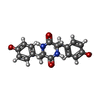 Database: PDB chemical components / ID: YTT Database: PDB chemical components / ID: YTT |
|---|---|
| Name | Name: ( |
-Chemical information
| Composition |  | ||||||||
|---|---|---|---|---|---|---|---|---|---|
| Others | Type: NON-POLYMER / PDB classification: HETAIN / Three letter code: YTT / Ideal coordinates details: Corina / Model coordinates PDB-ID: 3G5H | ||||||||
| History |
| ||||||||
 External links External links | UniChem / ChemSpider / BindingDB / Brenda / ChEBI / ChEMBL / CompTox / DrugBank / PubChem / SureChEMBL / Wikipedia search / Google search |
- Structure visualization
| Structure viewer | Molecule:  MolmilJmol/JSmol MolmilJmol/JSmol |
|---|
-Details
-SMILES
| ACDLabs 10.04 | | CACTVS 3.341 | OpenEye OEToolkits 1.5.0 | |
|---|
-SMILES CANONICAL
| CACTVS 3.341 | | OpenEye OEToolkits 1.5.0 | |
|---|
-InChI
| InChI 1.03 |
|---|
-InChIKey
| InChI 1.03 |
|---|
-SYSTEMATIC NAME
| ACDLabs 10.04 | (| OpenEye OEToolkits 1.5.0 | ( | |
|---|
-PDB entries
Showing all 5 items
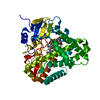
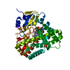
PDB-3g5h: 
Crystallographic analysis of cytochrome P450 cyp121
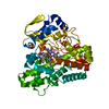
PDB-5wp2:
1.44 Angstrom crystal structure of CYP121 from Mycobacterium tuberculosis in complex with substrate and CN
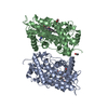
PDB-8q6y:
Crystal structure of Cytochrome P450 GymB5 from Streptomyces katrae in complex with cYY and Hypoxanthine

PDB-8tdp:
Time-resolved SFX-XFEL crystal structure of CYP121 bound with cYY reacted with peracetic acid for 200 milliseconds
PDB-8tdq:
SFX-XFEL structure of CYP121 cocrystallized with substrate cYY
