 Movie
Movie Controller


+ Open data

- Basic information
| Entry |  Database: PDB chemical components / ID: X90 Database: PDB chemical components / ID: X90 |
|---|---|
| Name | Name: |
-Chemical information
| Composition |  | ||||||
|---|---|---|---|---|---|---|---|
| Others | Type: NON-POLYMER / PDB classification: HETAIN / Three letter code: X90 / Ideal coordinates details: Corina / Model coordinates PDB-ID: 3OZW | ||||||
| History |
| ||||||
 External links External links | UniChem / Brenda / ChEBI / ChEMBL / ChemicalBook / CompTox / HMDB / LipidMaps / Metabolights / NMRShiftDB / Nikkaji / PubChem / PubChem_TPharma / SureChEMBL / ZINC / ChemSpider / Wikipedia search / Google search |

- Structure visualization
| Structure viewer | Molecule:  MolmilJmol/JSmol MolmilJmol/JSmol |
|---|
-Details
-SMILES
| ACDLabs 12.01 | | CACTVS 3.370 | OpenEye OEToolkits 1.7.0 | |
|---|
-SMILES CANONICAL
| CACTVS 3.370 | | OpenEye OEToolkits 1.7.0 | |
|---|
-InChI
| InChI 1.03 |
|---|
-InChIKey
| InChI 1.03 |
|---|
-SYSTEMATIC NAME
| ACDLabs 12.01 | | OpenEye OEToolkits 1.7.0 | |
|---|
-PDB entries
Showing all 4 items

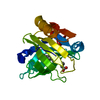
PDB-5uxy: 
The crystal structure of a DegV family protein from Eubacterium eligens loaded with heptadecanoic acid to 1.80 Angstrom resolution (ALTERNATIVE REFINEMENT OF PDB 3FDJ with HEPTADECANOIC acid)
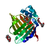
PDB-6gf9:
Thermodynamic, Crystallographic and Computational Studies of Non Mammalian Fatty Acid Binding to Bovine b-Lactoglobulin

PDB-6lq7:
Crystal Structure of E447A Acyl-CoA Dehydrogenase FadE5 mutant from Mycobacteria smegmatis in complex with C17CoA
PDB-7fdu:
The 0.86 angstrom X-ray structure of the human heart fatty acid-binding protein complexed with heptadecanoic acid
