 ムービー
ムービー コントローラー


+ データを開く

- 基本情報
基本情報
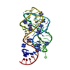
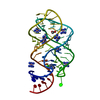
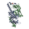
| 登録情報 |  データベース: PDB化学物質要素 / ID: 6GU データベース: PDB化学物質要素 / ID: 6GU |
|---|---|
| 名称 | 名称: |
-Chemical information
| 組成 |  | ||||||||
|---|---|---|---|---|---|---|---|---|---|
| その他 | タイプ: NON-POLYMER / PDB分類: HETAIN / 3文字コード: 6GU / 理論座標の詳細: Corina / モデル座標のPDB-ID: 3E9Z | ||||||||
| 履歴 |
| ||||||||
 外部リンク 外部リンク | UniChem / BindingDB / Brenda / ChEBI / ChEMBL / ChemicalBook / CompTox / HMDB / 日化辞 / PubChem / PubChem_TPharma / SureChEMBL / ZINC / ChemSpider / Wikipedia search / Google search |
- 構造の表示
構造の表示
| 構造ビューア | 分子:  MolmilJmol/JSmol MolmilJmol/JSmol |
|---|
-詳細
-SMILES
| ACDLabs 10.04 | | CACTVS 3.341 | OpenEye OEToolkits 1.5.0 | |
|---|
-SMILES CANONICAL
| CACTVS 3.341 | | OpenEye OEToolkits 1.5.0 | |
|---|
-InChI
| InChI 1.03 |
|---|
-InChIKey
| InChI 1.03 |
|---|
-SYSTEMATIC NAME
| ACDLabs 10.04 | | OpenEye OEToolkits 1.5.0 | |
|---|
-PDBエントリ
全6件を表示しています

PDB-3e9z: 
Crystal structure of purine nucleoside phosphorylase from Schistosoma mansoni in complex with 6-chloroguanine
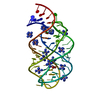
PDB-3fo4:
Crystal structure of guanine riboswitch C74U mutant bound to 6-chloroguanine
PDB-3ger:
Guanine riboswitch bound to 6-chloroguanine
PDB-3gog:
Guanine riboswitch A21G,U75C mutant bound to 6-chloroguanine

PDB-6q62:
Xanthomonas albilineans Dihydropteroate synthase in complex with (indole-2-carboxylic acid) and (6-chloroguanine)
PDB-6qoh:
Crystal structure of TrmD, a tRNA-(N1G37) methyltransferase, from Mycobacterium abscessus in complex with Fragment 11 (2-Amino-6-chloropurine)
