Mass: 1293.470 Da / Num. of mol.: 1 / Fragment: S2 Peptide, Residues 339-349 / Mutation: K340R, C346S Source method: isolated from a genetically manipulated source Source: (gene. exp.) Bos taurus (domestic cattle) / Gene: GNAT1 / Plasmid: GEV-S2 / Production host: Escherichia coli (E. coli) / References: UniProt: P04695
-
Experimental details
-
Experiment
Experiment
Method: SOLUTION NMR
NMR experiment
Conditions-ID
Experiment-ID
Solution-ID
Type
1
1
1
2D NOESY
1
2
1
2D 1H-15N HSQC (w/o 1H decoupling)
1
3
2
2D 1H-13C CT-HSQC (w/o 1H decoupling)
NMR details
Text: sample was studied in dark adapted state and after photo activation of rhodopsin by illuminating the sample for 60s with a focussed microscope light, chemical shifts of S2 peptide are identical ...Text: sample was studied in dark adapted state and after photo activation of rhodopsin by illuminating the sample for 60s with a focussed microscope light, chemical shifts of S2 peptide are identical in both states, TrNOEs and TrDCs are difference values between the dark and light-activated states. orientation of the bound peptide relative to the membrane normal was determined from residual dipolar couplings. the membrane normal that belongs to model 1 runs parallel to the y-axis of the coordinate frame in which the deposited s2 peptide coordinates are specified.
-
Sample preparation
Details
Solution-ID
Contents
Solvent system
1
2.6mM S2 peptide U-15N, 0.063mM rhodopsin as part of intact disk membranes from bovine retina; buffer: 10 mM HEPES, 20mM KCl, 0.05mM DTPA
90% H2O/10% D2O
2
2.6mM S2 peptide U-15N, 13C, 0.063mM rhodopsin as part of intact disk membranes from bovine retina; buffer: 10 mM HEPES, 20mM KCl, 0.05mM DTPA
90% H2O/10% D2O
Sample conditions
Ionic strength: 10 mM HEPES, 20 mM KCl / pH: 6.6 / Pressure: ambient / Temperature: 283 K
Crystal grow
*PLUS
Method: other / Details: NMR
-
NMR measurement
Radiation
Protocol: SINGLE WAVELENGTH / Monochromatic (M) / Laue (L): M
Radiation wavelength
Relative weight: 1
NMR spectrometer
Type
Manufacturer
Model
Field strength (MHz)
Spectrometer-ID
Bruker DMX
Bruker
DMX
750
1
Bruker DMX
Bruker
DMX
600
2
-
Processing
NMR software
Name
Version
Developer
Classification
NMRPipe
2.1
Delaglio
processing
X-PLOR
NIH
A.T. Brunger
structuresolution
X-PLOR
NIH
A.T. Brunger, N. Tjandra, C.D. Schwieters, J. Kuszewski, G.M. Clore
refinement
Refinement
Method: simulated annealing, molecular dynamics / Software ordinal: 1 Details: the structures are based on a total of 121 NOE-derived distance constraints, 12 NOE-derived dihedral angle restraints, and 38 residual dipolar couplings
NMR representative
Selection criteria: lowest energy structure
NMR ensemble
Conformer selection criteria: structures with the lowest energy Conformers calculated total number: 100 / Conformers submitted total number: 20
+
About Yorodumi
-
News
-
Feb 9, 2022. New format data for meta-information of EMDB entries
New format data for meta-information of EMDB entries
Version 3 of the EMDB header file is now the official format.
The previous official version 1.9 will be removed from the archive.
In the structure databanks used in Yorodumi, some data are registered as the other names, "COVID-19 virus" and "2019-nCoV". Here are the details of the virus and the list of structure data.
Jan 31, 2019. EMDB accession codes are about to change! (news from PDBe EMDB page)
EMDB accession codes are about to change! (news from PDBe EMDB page)
The allocation of 4 digits for EMDB accession codes will soon come to an end. Whilst these codes will remain in use, new EMDB accession codes will include an additional digit and will expand incrementally as the available range of codes is exhausted. The current 4-digit format prefixed with “EMD-” (i.e. EMD-XXXX) will advance to a 5-digit format (i.e. EMD-XXXXX), and so on. It is currently estimated that the 4-digit codes will be depleted around Spring 2019, at which point the 5-digit format will come into force.
The EM Navigator/Yorodumi systems omit the EMD- prefix.
Related info.:Q: What is EMD? / ID/Accession-code notation in Yorodumi/EM Navigator
Yorodumi is a browser for structure data from EMDB, PDB, SASBDB, etc.
This page is also the successor to EM Navigator detail page, and also detail information page/front-end page for Omokage search.
The word "yorodu" (or yorozu) is an old Japanese word meaning "ten thousand". "mi" (miru) is to see.
Related info.:EMDB / PDB / SASBDB / Comparison of 3 databanks / Yorodumi Search / Aug 31, 2016. New EM Navigator & Yorodumi / Yorodumi Papers / Jmol/JSmol / Function and homology information / Changes in new EM Navigator and Yorodumi
 Movie
Movie Controller

 Yorodumi
 Open data
 Basic information
 Components
 Keywords
 Function and homology information
 Authors
 Citation
 Structure visualization
 Downloads & links
 Other downloads
 PDBj
PDBj


 Assembly
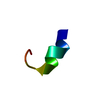

 HSQC
HSQC Sample preparation
 Processing