 Movie
Movie Controller


+ Open data

- Basic information
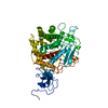
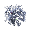
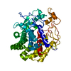
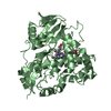
| Entry | Database: PDB / ID: 1ia6 | ||||||
|---|---|---|---|---|---|---|---|
| Title | CRYSTAL STRUCTURE OF THE CELLULASE CEL9M OF C. CELLULOLYTICUM | ||||||
 Components Components | CELLULASE CEL9M | ||||||
 Keywords Keywords | HYDROLASE / Cellullase / Alpha Barrel | ||||||
| Function / homology |  Function and homology information Function and homology informationHydrolases; Glycosylases; Glycosidases, i.e. enzymes that hydrolyse O- and S-glycosyl compounds / cellulase activity / cellulose catabolic process / metal ion binding Similarity search - Function | ||||||
| Biological species |  Clostridium cellulolyticum (bacteria) Clostridium cellulolyticum (bacteria) | ||||||
| Method |  X-RAY DIFFRACTION / MOLECULAR REPLACEMENT / Resolution: 1.8 Å X-RAY DIFFRACTION / MOLECULAR REPLACEMENT / Resolution: 1.8 Å | ||||||
 Authors Authors | Parsiegla, G. / Belaich, A. / Belaich, J.P. / Haser, R. | ||||||
 Citation Citation | Journal: Biochemistry / Year: 2002 Title: Crystal structure of the cellulase Cel9M enlightens structure/function relationships of the variable catalytic modules in glycoside hydrolases. Authors: Parsiegla, G. / Belaich, A. / Belaich, J.P. / Haser, R. | ||||||
| History |
|
- Structure visualization
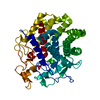
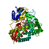
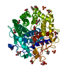
| Structure viewer | Molecule: MolmilJmol/JSmol |
|---|
- Downloads & links
-Download
| PDBx/mmCIF format | 1ia6.cif.gz | 104 KB | Display | PDBx/mmCIF format |
|---|---|---|---|---|
| PDB format | pdb1ia6.ent.gz | 77.7 KB | Display | PDB format |
| PDBx/mmJSON format | 1ia6.json.gz | Tree view | PDBx/mmJSON format | |
| Others |  Other downloads Other downloads |
-Validation report
| Arichive directory | https://data.pdbj.org/pub/pdb/validation_reports/ia/1ia6ftp://data.pdbj.org/pub/pdb/validation_reports/ia/1ia6 | HTTPS FTP |
|---|
-Related structure data
| Related structure data |  1ia7C 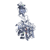 1js4S S: Starting model for refinement C: citing same article ( |
|---|---|
| Similar structure data |


-Links
 PDBj
PDBj
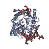
- Assembly
| Deposited unit | 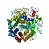
| ||||||||
|---|---|---|---|---|---|---|---|---|---|
| 1 |
| ||||||||
| Unit cell |
|
-Components
-Protein , 1 types, 1 molecules A
| #1: Protein | Mass: 48697.875 Da / Num. of mol.: 1 / Fragment: CATALYTIC MODULE Source method: isolated from a genetically manipulated source Source: (gene. exp.) Clostridium cellulolyticum (bacteria) / Production host: |
|---|
-Non-polymers , 5 types, 266 molecules 


| #2: Chemical | ChemComp-CA /  Mass: 40.078 Da / Num. of mol.: 1 / Source method: obtained synthetically / Formula: Ca Mass: 40.078 Da / Num. of mol.: 1 / Source method: obtained synthetically / Formula: Ca |
|---|---|
| #3: Chemical | ChemComp-ZN /  Mass: 65.409 Da / Num. of mol.: 1 / Source method: obtained synthetically / Formula: Zn Mass: 65.409 Da / Num. of mol.: 1 / Source method: obtained synthetically / Formula: Zn |
| #4: Chemical | ChemComp-NI /  Mass: 58.693 Da / Num. of mol.: 1 / Source method: obtained synthetically / Formula: Ni Mass: 58.693 Da / Num. of mol.: 1 / Source method: obtained synthetically / Formula: Ni |
| #5: Chemical | ChemComp-SO4 /  Mass: 96.063 Da / Num. of mol.: 1 / Source method: obtained synthetically / Formula: SO4 Mass: 96.063 Da / Num. of mol.: 1 / Source method: obtained synthetically / Formula: SO4 |
| #6: Water | ChemComp-HOH / Mass: 18.015 Da / Num. of mol.: 262 / Source method: isolated from a natural source / Formula: H2O |
-Experimental details
-Experiment
| Experiment | Method: X-RAY DIFFRACTION / Number of used crystals: 1 |
|---|
- Sample preparation
| Crystal | Density Matthews: 1.93 Å3/Da / Density % sol: 36.4 % | |||||||||||||||||||||||||||||||||||
|---|---|---|---|---|---|---|---|---|---|---|---|---|---|---|---|---|---|---|---|---|---|---|---|---|---|---|---|---|---|---|---|---|---|---|---|---|
| Crystal grow | Temperature: 290 K / Method: vapor diffusion, hanging drop / pH: 7.5 Details: ammonium sulphate, calcium chloride, HEPES, pH 7.5, VAPOR DIFFUSION, HANGING DROP, temperature 290K | |||||||||||||||||||||||||||||||||||
| Crystal grow | *PLUS Temperature: 18 ℃ | |||||||||||||||||||||||||||||||||||
| Components of the solutions | *PLUS
|
-Data collection
| Diffraction | Mean temperature: 290 K |
|---|---|
| Diffraction source | Source: ROTATING ANODE / Type: ENRAF-NONIUS FU581 / Wavelength: 1.5418 Å |
| Detector | Type: MARRESEARCH / Detector: IMAGE PLATE / Date: Oct 14, 2000 / Details: Osmic confocal mirrors |
| Radiation | Monochromator: confocal mirrors / Protocol: SINGLE WAVELENGTH / Monochromatic (M) / Laue (L): M / Scattering type: x-ray |
| Radiation wavelength | Wavelength: 1.5418 Å / Relative weight: 1 |
| Reflection | Resolution: 1.8→16 Å / Num. all: 170027 / Num. obs: 33837 / % possible obs: 97.8 % / Observed criterion σ(F): 0 / Observed criterion σ(I): 0 / Redundancy: 5 % / Biso Wilson estimate: 14.8 Å2 / Rmerge(I) obs: 0.083 / Rsym value: 6.6 / Net I/σ(I): 9 |
| Reflection shell | Resolution: 1.8→1.86 Å / Redundancy: 3 % / Rmerge(I) obs: 0.295 / Mean I/σ(I) obs: 2.9 / Num. unique all: 9028 / Rsym value: 23 / % possible all: 88.7 |
| Reflection | *PLUS Lowest resolution: 30 Å / Num. measured all: 170027 / Rmerge(I) obs: 0.083 |
| Reflection shell | *PLUS % possible obs: 88.7 % / Rmerge(I) obs: 0.295 |
- Processing
| Software |
| ||||||||||||||||||||||||||||||||||||||||||||||||||||||||||||||||||||||||||||||||
|---|---|---|---|---|---|---|---|---|---|---|---|---|---|---|---|---|---|---|---|---|---|---|---|---|---|---|---|---|---|---|---|---|---|---|---|---|---|---|---|---|---|---|---|---|---|---|---|---|---|---|---|---|---|---|---|---|---|---|---|---|---|---|---|---|---|---|---|---|---|---|---|---|---|---|---|---|---|---|---|---|---|
| Refinement | Method to determine structure: MOLECULAR REPLACEMENT Starting model: Model with Cel9M sequence based on PDB ENTRY 1JS4. Resolution: 1.8→29.24 Å / Rfactor Rfree error: 0.005 / Data cutoff high absF: 1252803.33 / Data cutoff low absF: 0 / Isotropic thermal model: RESTRAINED / Cross valid method: THROUGHOUT / σ(F): 0 / σ(I): 0 / Stereochemistry target values: Engh & Huber Details: The sequence in two flexible loops between L239 and N246 or A373 and D376 could not be fitted and is omitted. Mass spectrometric analysis has indicated that the sequence GTIVNPPVKK should ...Details: The sequence in two flexible loops between L239 and N246 or A373 and D376 could not be fitted and is omitted. Mass spectrometric analysis has indicated that the sequence GTIVNPPVKK should additionally be present at the C-terminus in the crystal, but it could not be traced in the electron density.
| ||||||||||||||||||||||||||||||||||||||||||||||||||||||||||||||||||||||||||||||||
| Solvent computation | Solvent model: FLAT MODEL / Bsol: 49.21 Å2 / ksol: 0.377 e/Å3 | ||||||||||||||||||||||||||||||||||||||||||||||||||||||||||||||||||||||||||||||||
| Displacement parameters | Biso mean: 16.3 Å2
| ||||||||||||||||||||||||||||||||||||||||||||||||||||||||||||||||||||||||||||||||
| Refine analyze |
| ||||||||||||||||||||||||||||||||||||||||||||||||||||||||||||||||||||||||||||||||
| Refinement step | Cycle: LAST / Resolution: 1.8→29.24 Å
| ||||||||||||||||||||||||||||||||||||||||||||||||||||||||||||||||||||||||||||||||
| Refine LS restraints |
| ||||||||||||||||||||||||||||||||||||||||||||||||||||||||||||||||||||||||||||||||
| LS refinement shell | Resolution: 1.8→1.91 Å / Rfactor Rfree error: 0.014 / Total num. of bins used: 6
| ||||||||||||||||||||||||||||||||||||||||||||||||||||||||||||||||||||||||||||||||
| Xplor file |
| ||||||||||||||||||||||||||||||||||||||||||||||||||||||||||||||||||||||||||||||||
| Refinement | *PLUS Lowest resolution: 30 Å / Rfactor Rfree: 0.189 / Rfactor Rwork: 0.158 | ||||||||||||||||||||||||||||||||||||||||||||||||||||||||||||||||||||||||||||||||
| Solvent computation | *PLUS | ||||||||||||||||||||||||||||||||||||||||||||||||||||||||||||||||||||||||||||||||
| Displacement parameters | *PLUS | ||||||||||||||||||||||||||||||||||||||||||||||||||||||||||||||||||||||||||||||||
| Refine LS restraints | *PLUS Type: c_angle_deg / Dev ideal: 1.2 | ||||||||||||||||||||||||||||||||||||||||||||||||||||||||||||||||||||||||||||||||
| LS refinement shell | *PLUS Lowest resolution: 1.86 Å / Rfactor Rfree: 0.249 / Rfactor Rwork: 0.222 |
