 Movie
Movie Controller

[English] 日本語
 Yorodumi
- PDB-1h41: Pseudomonas cellulosa E292A alpha-D-glucuronidase mutant complexe... -
+ Open data

- Basic information
| Entry | Database: PDB / ID: 1h41 | ||||||
|---|---|---|---|---|---|---|---|
| Title | Pseudomonas cellulosa E292A alpha-D-glucuronidase mutant complexed with aldotriuronic acid | ||||||
 Components Components | ALPHA-GLUCURONIDASE | ||||||
 Keywords Keywords | HYDROLASE / GLUCURONIDASE / (ALPHA-BETA)8 BARREL / GLYCOSIDE HYDROLASE / GLUCURONIC ACID | ||||||
| Function / homology |  Function and homology information Function and homology informationxylan alpha-1,2-glucuronosidase / xylan alpha-1,2-glucuronosidase activity / alpha-glucuronidase activity / glucuronoxylan catabolic process / Hydrolases; Glycosylases; Glycosidases, i.e. enzymes that hydrolyse O- and S-glycosyl compounds / cell outer membrane / extracellular region Similarity search - Function | ||||||
| Biological species |  PSEUDOMONAS CELLULOSA (bacteria) PSEUDOMONAS CELLULOSA (bacteria) | ||||||
| Method |  X-RAY DIFFRACTION / SYNCHROTRON / MOLECULAR REPLACEMENT / Resolution: 1.5 Å X-RAY DIFFRACTION / SYNCHROTRON / MOLECULAR REPLACEMENT / Resolution: 1.5 Å | ||||||
 Authors Authors | Nurizzo, D. / Nagy, T. / Gilbert, H.J. / Davies, G.J. | ||||||
 Citation Citation | Journal: J.Biol.Chem. / Year: 2003 Title: The Alpha-Glucuronidase,Glca67A,of Cellvibrio Japonicus Utilizes the Carboxylate and Methyl Groups of Aldobiouronic Acid as Important Substrate Recognition Determinants Authors: Nagy, T. / Nurizzo, D. / Davies, G.J. / Biely, P. / Lakey, J.H. / Bolam, D.N. / Gilbert, H.J. | ||||||
| History |
| ||||||
| Remark 700 | SHEET DETERMINATION METHOD: DSSP THE SHEETS PRESENTED AS "AB" AND "BB" IN EACH CHAIN ON SHEET ... SHEET DETERMINATION METHOD: DSSP THE SHEETS PRESENTED AS "AB" AND "BB" IN EACH CHAIN ON SHEET RECORDS BELOW IS ACTUALLY AN 8-STRANDED BARREL THIS IS REPRESENTED BY A 9-STRANDED SHEET IN WHICH THE FIRST AND LAST STRANDS ARE IDENTICAL. |
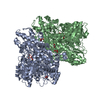
- Structure visualization
| Structure viewer | Molecule: MolmilJmol/JSmol |
|---|
- Downloads & links
-Download
| PDBx/mmCIF format | 1h41.cif.gz | 327.8 KB | Display | PDBx/mmCIF format |
|---|---|---|---|---|
| PDB format | pdb1h41.ent.gz | 263.4 KB | Display | PDB format |
| PDBx/mmJSON format | 1h41.json.gz | Tree view | PDBx/mmJSON format | |
| Others |  Other downloads Other downloads |
-Validation report
| Arichive directory | https://data.pdbj.org/pub/pdb/validation_reports/h4/1h41ftp://data.pdbj.org/pub/pdb/validation_reports/h4/1h41 | HTTPS FTP |
|---|
-Related structure data
| Related structure data | 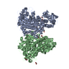 1gqiS S: Starting model for refinement |
|---|---|
| Similar structure data |


-Links
 PDBj
PDBj

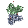
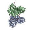
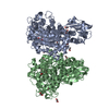
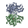
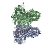
- Assembly
| Deposited unit | 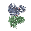
| ||||||||
|---|---|---|---|---|---|---|---|---|---|
| 1 |
| ||||||||
| Unit cell |
|
-Components
| #1: Protein | Mass: 80381.562 Da / Num. of mol.: 2 / Mutation: YES Source method: isolated from a genetically manipulated source Source: (gene. exp.) PSEUDOMONAS CELLULOSA (bacteria) / Plasmid: PTN1 / Production host: References: UniProt: Q8VP74, UniProt: B3PC73*PLUS, EC: 3.2.1.139 #2: Sugar | 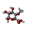  Type: D-saccharide, alpha linking / Mass: 208.166 Da / Num. of mol.: 2 Type: D-saccharide, alpha linking / Mass: 208.166 Da / Num. of mol.: 2Source method: isolated from a genetically manipulated source Formula: C7H12O7 #3: Chemical | ChemComp-EDO /   Mass: 62.068 Da / Num. of mol.: 28 / Source method: obtained synthetically / Formula: C2H6O2 Mass: 62.068 Da / Num. of mol.: 28 / Source method: obtained synthetically / Formula: C2H6O2#4: Chemical | ChemComp-CO /   Mass: 58.933 Da / Num. of mol.: 10 / Source method: obtained synthetically / Formula: Co Mass: 58.933 Da / Num. of mol.: 10 / Source method: obtained synthetically / Formula: Co#5: Water | ChemComp-HOH / |  Mass: 18.015 Da / Num. of mol.: 1667 / Source method: isolated from a natural source / Formula: H2O Mass: 18.015 Da / Num. of mol.: 1667 / Source method: isolated from a natural source / Formula: H2OCompound details | ENGINEERED | |
|---|
-Experimental details
-Experiment
| Experiment | Method: X-RAY DIFFRACTION / Number of used crystals: 1 |
|---|
- Sample preparation
| Crystal | Density Matthews: 2.4 Å3/Da / Density % sol: 48.5 % | ||||||||||||||||||||||||||||||||||||||||||
|---|---|---|---|---|---|---|---|---|---|---|---|---|---|---|---|---|---|---|---|---|---|---|---|---|---|---|---|---|---|---|---|---|---|---|---|---|---|---|---|---|---|---|---|
| Crystal grow | pH: 8 Details: 15% PEG3350, 250MM MGCL2, 5MM TRIS-HCL PH8.0, 20% GLYCEROL, pH 8.00 | ||||||||||||||||||||||||||||||||||||||||||
| Crystal grow | *PLUS Temperature: 20 ℃ / Method: vapor diffusion, hanging drop / Details: Nurizzo, D., (2002) Structure, 10, 547. | ||||||||||||||||||||||||||||||||||||||||||
| Components of the solutions | *PLUS
|
-Data collection
| Diffraction | Mean temperature: 100 K |
|---|---|
| Diffraction source | Source: SYNCHROTRON / Site: ESRF  / Beamline: ID14-4 / Wavelength: 0.9392 / Beamline: ID14-4 / Wavelength: 0.9392 |
| Detector | Type: ADSC CCD / Detector: CCD / Date: Dec 15, 2001 |
| Radiation | Protocol: SINGLE WAVELENGTH / Monochromatic (M) / Laue (L): M / Scattering type: x-ray |
| Radiation wavelength | Wavelength: 0.9392 Å / Relative weight: 1 |
| Reflection | Resolution: 1.5→20 Å / Num. obs: 212953 / % possible obs: 91.2 % / Redundancy: 2.7 % / Rmerge(I) obs: 0.041 / Net I/σ(I): 20.06 |
| Reflection shell | Resolution: 1.5→1.53 Å / Redundancy: 2.5 % / Rmerge(I) obs: 0.215 / Mean I/σ(I) obs: 4.23 / % possible all: 52.9 |
| Reflection | *PLUS Highest resolution: 1.5 Å / Lowest resolution: 20 Å / Rmerge(I) obs: 0.047 |
| Reflection shell | *PLUS % possible obs: 52.9 % / Redundancy: 2.6 % / Num. unique obs: 8276 / Rmerge(I) obs: 0.237 / Mean I/σ(I) obs: 4.2 |
- Processing
| Software |
| ||||||||||||||||||||||||||||||||||||||||||||||||||||||||||||||||||||||||||||||||||||||||||||||||||||||||||||||||||||||||||||||||||||||||||||||||||||||||||||||||||||||||||||||||||||||
|---|---|---|---|---|---|---|---|---|---|---|---|---|---|---|---|---|---|---|---|---|---|---|---|---|---|---|---|---|---|---|---|---|---|---|---|---|---|---|---|---|---|---|---|---|---|---|---|---|---|---|---|---|---|---|---|---|---|---|---|---|---|---|---|---|---|---|---|---|---|---|---|---|---|---|---|---|---|---|---|---|---|---|---|---|---|---|---|---|---|---|---|---|---|---|---|---|---|---|---|---|---|---|---|---|---|---|---|---|---|---|---|---|---|---|---|---|---|---|---|---|---|---|---|---|---|---|---|---|---|---|---|---|---|---|---|---|---|---|---|---|---|---|---|---|---|---|---|---|---|---|---|---|---|---|---|---|---|---|---|---|---|---|---|---|---|---|---|---|---|---|---|---|---|---|---|---|---|---|---|---|---|---|---|
| Refinement | Method to determine structure: MOLECULAR REPLACEMENT Starting model: PDB ENTRY 1GQI Resolution: 1.5→20 Å / Cor.coef. Fo:Fc: 0.978 / Cor.coef. Fo:Fc free: 0.968 / SU B: 0.943 / SU ML: 0.035 / Cross valid method: THROUGHOUT / ESU R: 0.069 / ESU R Free: 0.059 / Stereochemistry target values: MAXIMUM LIKELIHOOD / Details: HYDROGENS HAVE BEEN ADDED IN THE RIDING POSITIONS
| ||||||||||||||||||||||||||||||||||||||||||||||||||||||||||||||||||||||||||||||||||||||||||||||||||||||||||||||||||||||||||||||||||||||||||||||||||||||||||||||||||||||||||||||||||||||
| Solvent computation | Ion probe radii: 0.8 Å / Shrinkage radii: 0.8 Å / VDW probe radii: 1.4 Å / Solvent model: BABINET MODEL WITH MASK | ||||||||||||||||||||||||||||||||||||||||||||||||||||||||||||||||||||||||||||||||||||||||||||||||||||||||||||||||||||||||||||||||||||||||||||||||||||||||||||||||||||||||||||||||||||||
| Displacement parameters | Biso mean: 12.76 Å2
| ||||||||||||||||||||||||||||||||||||||||||||||||||||||||||||||||||||||||||||||||||||||||||||||||||||||||||||||||||||||||||||||||||||||||||||||||||||||||||||||||||||||||||||||||||||||
| Refinement step | Cycle: LAST / Resolution: 1.5→20 Å
| ||||||||||||||||||||||||||||||||||||||||||||||||||||||||||||||||||||||||||||||||||||||||||||||||||||||||||||||||||||||||||||||||||||||||||||||||||||||||||||||||||||||||||||||||||||||
| Refine LS restraints |
|