 Movie
Movie Controller


+ Open data

- Basic information
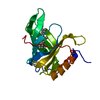
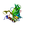
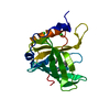
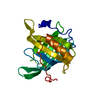
| Entry | Database: PDB / ID: 1aqb | ||||||
|---|---|---|---|---|---|---|---|
| Title | RETINOL-BINDING PROTEIN (RBP) FROM PIG PLASMA | ||||||
 Components Components | RETINOL-BINDING PROTEIN | ||||||
 Keywords Keywords | RETINOL TRANSPORT / RETINOIDS / VITAMIN A / CADMIUM ION | ||||||
| Function / homology |  Function and homology information Function and homology informationretinol transport / retinol transmembrane transporter activity / retinal binding / retinol binding / : Similarity search - Function | ||||||
| Biological species |  | ||||||
| Method |  X-RAY DIFFRACTION / MOLECULAR REPLACEMENT / Resolution: 1.65 Å X-RAY DIFFRACTION / MOLECULAR REPLACEMENT / Resolution: 1.65 Å | ||||||
 Authors Authors | Zanotti, G. / Panzalorto, M. / Marcato, A. / Malpeli, G. / Folli, C. / Berni, R. | ||||||
 Citation Citation | Journal: Acta Crystallogr.,Sect.D / Year: 1998 Title: Structure of pig plasma retinol-binding protein at 1.65 A resolution. Authors: Zanotti, G. / Panzalorto, M. / Marcato, A. / Malpeli, G. / Folli, C. / Berni, R. #1: Journal: J.Biol.Chem. / Year: 1993Title: Crystal Structure of Liganded and Unliganded Forms of Bovine Plasma Retinol-Binding Protein Authors: Zanotti, G. / Berni, R. / Monaco, H.L. #2: Journal: J.Mol.Biol. / Year: 1993Title: Crystal Structure of the Trigonal Form of Human Plasma Retinol-Binding Protein at 2.5 A Resolution Authors: Zanotti, G. / Ottonello, S. / Berni, R. / Monaco, H.L. | ||||||
| History |
|
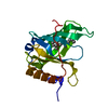
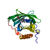
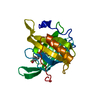
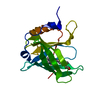
- Structure visualization
| Structure viewer | Molecule: MolmilJmol/JSmol |
|---|
- Downloads & links
-Download
| PDBx/mmCIF format | 1aqb.cif.gz | 53.1 KB | Display | PDBx/mmCIF format |
|---|---|---|---|---|
| PDB format | pdb1aqb.ent.gz | 36.9 KB | Display | PDB format |
| PDBx/mmJSON format | 1aqb.json.gz | Tree view | PDBx/mmJSON format | |
| Others |  Other downloads Other downloads |
-Validation report
| Arichive directory | https://data.pdbj.org/pub/pdb/validation_reports/aq/1aqbftp://data.pdbj.org/pub/pdb/validation_reports/aq/1aqb | HTTPS FTP |
|---|
-Related structure data
| Related structure data |  1hbpS S: Starting model for refinement |
|---|---|
| Similar structure data |


-Links
 PDBj
PDBj

- Assembly
| Deposited unit | 
| ||||||||
|---|---|---|---|---|---|---|---|---|---|
| 1 |
| ||||||||
| Unit cell |
|
-Components
| #1: Protein | Mass: 21183.656 Da / Num. of mol.: 1 / Source method: isolated from a natural source / Source: (natural) |
|---|---|
| #2: Chemical | ChemComp-CD /   Mass: 112.411 Da / Num. of mol.: 1 / Source method: obtained synthetically / Formula: Cd Mass: 112.411 Da / Num. of mol.: 1 / Source method: obtained synthetically / Formula: Cd |
| #3: Chemical | ChemComp-RTL /   Mass: 286.452 Da / Num. of mol.: 1 / Source method: obtained synthetically / Formula: C20H30O Mass: 286.452 Da / Num. of mol.: 1 / Source method: obtained synthetically / Formula: C20H30O |
| #4: Water | ChemComp-HOH /  Mass: 18.015 Da / Num. of mol.: 124 / Source method: isolated from a natural source / Formula: H2O Mass: 18.015 Da / Num. of mol.: 124 / Source method: isolated from a natural source / Formula: H2O |
| Has protein modification | Y |
-Experimental details
-Experiment
| Experiment | Method: X-RAY DIFFRACTION / Number of used crystals: 1 |
|---|
- Sample preparation
| Crystal | Density Matthews: 2.1 Å3/Da / Density % sol: 41 % | |||||||||||||||||||||||||
|---|---|---|---|---|---|---|---|---|---|---|---|---|---|---|---|---|---|---|---|---|---|---|---|---|---|---|
| Crystal grow | Method: vapor diffusion, sitting drop / pH: 6.8 Details: SITTING DROP VAPOR DIFFUSION METHOD, AT A FINAL PROTEIN CONCENTRATION OF 8 MG/ML IN THE PRESENCE OF 8% 2-METHYL-2,4-PENTANEDIOL, 3 MM CADMIUM ACETATE, 0.1 M TRIS-ACETATE, PH=6.8, vapor diffusion - sitting drop | |||||||||||||||||||||||||
| Crystal grow | *PLUS Temperature: 277 K / Method: vapor diffusion, sitting drop | |||||||||||||||||||||||||
| Components of the solutions | *PLUS
|
-Data collection
| Diffraction | Mean temperature: 300 K |
|---|---|
| Diffraction source | Source: ROTATING ANODE / Type: MACSCIENCE M18X / Wavelength: 1.5418 |
| Detector | Type: SIEMENS / Detector: AREA DETECTOR / Date: Sep 1, 1996 |
| Radiation | Monochromator: GRAPHITE(002) / Monochromatic (M) / Laue (L): M / Scattering type: x-ray |
| Radiation wavelength | Wavelength: 1.5418 Å / Relative weight: 1 |
| Reflection | Resolution: 1.65→55 Å / Num. obs: 20127 / % possible obs: 91 % / Observed criterion σ(I): 0 / Redundancy: 4.2 % / Rmerge(I) obs: 0.064 / Rsym value: 0.064 / Net I/σ(I): 8 |
| Reflection shell | Resolution: 1.65→1.72 Å / Redundancy: 2.5 % / Rmerge(I) obs: 0.19 / Mean I/σ(I) obs: 4 / Rsym value: 0.19 / % possible all: 56 |
| Reflection | *PLUS Num. measured all: 84968 |
- Processing
| Software |
| |||||||||||||||||||||||||||||||||
|---|---|---|---|---|---|---|---|---|---|---|---|---|---|---|---|---|---|---|---|---|---|---|---|---|---|---|---|---|---|---|---|---|---|---|
| Refinement | Method to determine structure: MOLECULAR REPLACEMENT Starting model: 1HBP Resolution: 1.65→55 Å / Num. parameters: 6308 / Num. restraintsaints: 5678 / Cross valid method: FREE R-VALUE / σ(F): 0 / Stereochemistry target values: ENGH AND HUBER Details: NO RESTRAINTS WERE APPLIED ON THE PLANARITY OF RETINOL MOLECULE
| |||||||||||||||||||||||||||||||||
| Solvent computation | Solvent model: MOEWS & KRETSINGER | |||||||||||||||||||||||||||||||||
| Refine analyze | Num. disordered residues: 3 / Occupancy sum hydrogen: 5272 / Occupancy sum non hydrogen: 6284 | |||||||||||||||||||||||||||||||||
| Refinement step | Cycle: LAST / Resolution: 1.65→55 Å
| |||||||||||||||||||||||||||||||||
| Refine LS restraints |
|
