 Movie
Movie Controller


+ Open data

- Basic information
| Entry | Database: EMDB / ID: EMD-9212 | |||||||||
|---|---|---|---|---|---|---|---|---|---|---|
| Title | 12-meric ClyA pore complex | |||||||||
 Map data Map data | Dodecamer | |||||||||
 Sample Sample |
| |||||||||
 Keywords Keywords | Pore-forming toxin / MEMBRANE PROTEIN / Toxin | |||||||||
| Function / homology |  Function and homology information Function and homology informationhemolysis in another organism / Secretion of toxins / toxin activity / periplasmic space / host cell plasma membrane / extracellular region / identical protein binding Similarity search - Function | |||||||||
| Biological species |  | |||||||||
| Method | single particle reconstruction / cryo EM / Resolution: 2.8 Å | |||||||||
 Authors Authors | Peng W / de Souza Santos M | |||||||||
 Citation Citation | Journal: PLoS One / Year: 2019 Title: High-resolution cryo-EM structures of the E. coli hemolysin ClyA oligomers. Authors: Wei Peng / Marcela de Souza Santos / Yang Li / Diana R Tomchick / Kim Orth /  Abstract: Pore-forming proteins (PFPs) represent a functionally important protein family, that are found in organisms from viruses to humans. As a major branch of PFPs, bacteria pore-forming toxins (PFTs) ...Pore-forming proteins (PFPs) represent a functionally important protein family, that are found in organisms from viruses to humans. As a major branch of PFPs, bacteria pore-forming toxins (PFTs) permeabilize membranes and usually cause the death of target cells. E. coli hemolysin ClyA is the first member with the pore complex structure solved among α-PFTs, employing α-helices as transmembrane elements. ClyA is proposed to form pores composed of various numbers of protomers. With high-resolution cryo-EM structures, we observe that ClyA pore complexes can exist as newly confirmed oligomers of a tridecamer and a tetradecamer, at estimated resolutions of 3.2 Å and 4.3 Å, respectively. The 2.8 Å cryo-EM structure of a dodecamer dramatically improves the existing structural model. Structural analysis indicates that protomers from distinct oligomers resemble each other and neighboring protomers adopt a conserved interaction mode. We also show a stabilized intermediate state of ClyA during the transition process from soluble monomers to pore complexes. Unexpectedly, even without the formation of mature pore complexes, ClyA can permeabilize membranes and allow leakage of particles less than ~400 Daltons. In addition, we are the first to show that ClyA forms pore complexes in the presence of cholesterol within artificial liposomes. These findings provide new mechanistic insights into the dynamic process of pore assembly for the prototypical α-PFT ClyA. | |||||||||
| History |
|
- Structure visualization
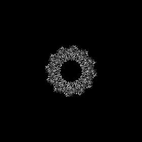
| Movie |
Movie viewer |
|---|---|
| Structure viewer | EM map: SurfViewMolmilJmol/JSmol |
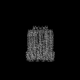
| Supplemental images |

- Downloads & links
-EMDB archive
| Map data | emd_9212.map.gz | 77.9 MB | EMDB map data format | |
|---|---|---|---|---|
| Header (meta data) | emd-9212-v30.xmlemd-9212.xml | 8.8 KB 8.8 KB | Display Display | EMDB header |
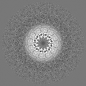
| Images | 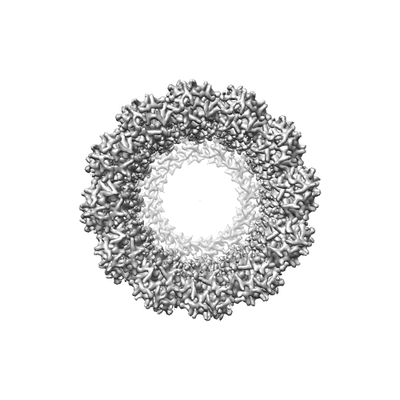 emd_9212.png emd_9212.png | 40.1 KB | ||
| Filedesc metadata | emd-9212.cif.gz | 5.2 KB | ||
| Archive directory |  http://ftp.pdbj.org/pub/emdb/structures/EMD-9212ftp://ftp.pdbj.org/pub/emdb/structures/EMD-9212 http://ftp.pdbj.org/pub/emdb/structures/EMD-9212ftp://ftp.pdbj.org/pub/emdb/structures/EMD-9212 | HTTPS FTP |
-Related structure data
| Related structure data |  6mrtMC  9213C  9214C  6mruC  6mrwC M: atomic model generated by this map C: citing same article ( |
|---|---|
| Similar structure data |


-Links
| EMDB pages | EMDB (EBI/PDBe) / EMDataResource |
|---|
-Map
| File | Download / File: emd_9212.map.gz / Format: CCP4 / Size: 83.7 MB / Type: IMAGE STORED AS FLOATING POINT NUMBER (4 BYTES) | ||||||||||||||||||||||||||||||||||||||||||||||||||||||||||||||||||||
|---|---|---|---|---|---|---|---|---|---|---|---|---|---|---|---|---|---|---|---|---|---|---|---|---|---|---|---|---|---|---|---|---|---|---|---|---|---|---|---|---|---|---|---|---|---|---|---|---|---|---|---|---|---|---|---|---|---|---|---|---|---|---|---|---|---|---|---|---|---|
| Annotation | Dodecamer | ||||||||||||||||||||||||||||||||||||||||||||||||||||||||||||||||||||
| Projections & slices | Image control
Images are generated by Spider. | ||||||||||||||||||||||||||||||||||||||||||||||||||||||||||||||||||||
| Voxel size | X=Y=Z: 1.07 Å | ||||||||||||||||||||||||||||||||||||||||||||||||||||||||||||||||||||
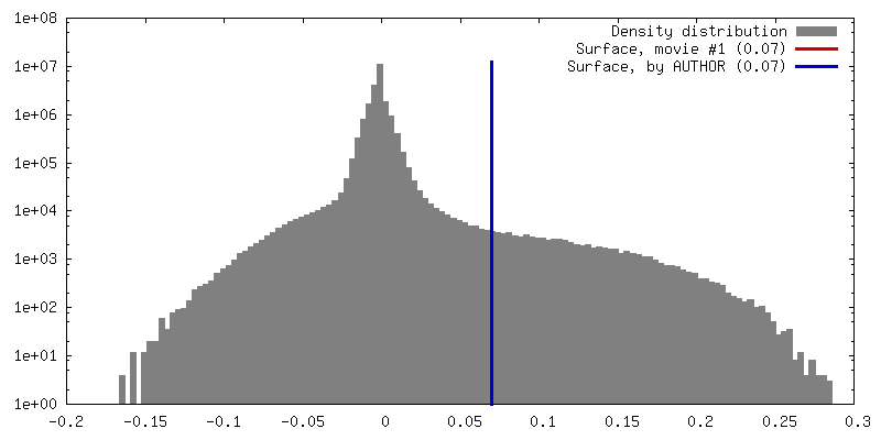
| Density |
| ||||||||||||||||||||||||||||||||||||||||||||||||||||||||||||||||||||
| Symmetry | Space group: 1 | ||||||||||||||||||||||||||||||||||||||||||||||||||||||||||||||||||||
| Details | EMDB XML:
CCP4 map header:
| ||||||||||||||||||||||||||||||||||||||||||||||||||||||||||||||||||||
 Z (Sec.)
 Y (Row.)
 X (Col.)


-Supplemental data
- Sample components
-Entire : 12-meric ClyA pore complex
| Entire | Name: 12-meric ClyA pore complex |
|---|---|
| Components |
|
-Supramolecule #1: 12-meric ClyA pore complex
| Supramolecule | Name: 12-meric ClyA pore complex / type: complex / ID: 1 / Parent: 0 / Macromolecule list: all |
|---|---|
| Source (natural) | Organism: |
| Molecular weight | Theoretical: 410 KDa |
-Macromolecule #1: Hemolysin E, chromosomal
| Macromolecule | Name: Hemolysin E, chromosomal / type: protein_or_peptide / ID: 1 / Number of copies: 12 / Enantiomer: LEVO |
|---|---|
| Source (natural) | Organism: |
| Molecular weight | Theoretical: 36.131816 KDa |
| Recombinant expression | Organism: |
| Sequence | String: MGSSHHHHHH SQDLDEVDAG SMTEIVADKT VEVVKNAIET ADGALDLYNK YLDQVIPWQT FDETIKELSR FKQEYSQAAS VLVGDIKTL LMDSQDKYFE ATQTVYEWCG VATQLLAAYI LLFDEYNEKK ASAQKDILIK VLDDGITKLN EAQKSLLVSS Q SFNNASGK ...String: MGSSHHHHHH SQDLDEVDAG SMTEIVADKT VEVVKNAIET ADGALDLYNK YLDQVIPWQT FDETIKELSR FKQEYSQAAS VLVGDIKTL LMDSQDKYFE ATQTVYEWCG VATQLLAAYI LLFDEYNEKK ASAQKDILIK VLDDGITKLN EAQKSLLVSS Q SFNNASGK LLALDSQLTN DFSEKSSYFQ SQVDKIRKEA YAGAAAGVVA GPFGLIISYS IAAGVVEGKL IPELKNKLKS VQ NFFTTLS NTVKQANKDI DAAKLKLTTE IVAIGEIKTE TETTRFYVDY DDLMLSLLKE AAKKMINTCN EYQKRHGKKT LFE VPEV UniProtKB: Hemolysin E, chromosomal |
-Experimental details
-Structure determination
| Method | cryo EM |
|---|---|
 Processing Processing | single particle reconstruction |
| Aggregation state | particle |
-Sample preparation
| Buffer | pH: 8 |
|---|---|
| Grid | Details: unspecified |
| Vitrification | Cryogen name: ETHANE |
- Electron microscopy
| Microscope | FEI TITAN KRIOS |
|---|---|
| Image recording | Film or detector model: GATAN K2 SUMMIT (4k x 4k) / Detector mode: SUPER-RESOLUTION / Average electron dose: 50.0 e/Å2 |
| Electron beam | Acceleration voltage: 300 kV / Electron source:  FIELD EMISSION GUN FIELD EMISSION GUN |
| Electron optics | Illumination mode: FLOOD BEAM / Imaging mode: BRIGHT FIELD |
| Experimental equipment |  Model: Titan Krios / Image courtesy: FEI Company |
-Image processing
| Startup model | Type of model: PDB ENTRY |
|---|---|
| Final reconstruction | Resolution.type: BY AUTHOR / Resolution: 2.8 Å / Resolution method: FSC 0.143 CUT-OFF / Number images used: 482946 |
| Initial angle assignment | Type: NOT APPLICABLE |
| Final angle assignment | Type: NOT APPLICABLE |
