[English] 日本語
Yorodumi
- PDB-8gcy: Co-crystal structure of CBL-B in complex with N-Aryl isoindolin-1... -

+
Open data


ID or keywords:

Loading...

-
Basic information

Entry
Database: PDB / ID: 8gcy
TitleCo-crystal structure of CBL-B in complex with N-Aryl isoindolin-1-one inhibitor
ComponentsE3 ubiquitin-protein ligase CBL-B
KeywordsLIGASE/LIGASE INHIBITOR / E3 ligase / CBL-B / inhibitor / SGC / LIGASE / LIGASE-LIGASE INHIBITOR complex
Function / homology
Function and homology information


regulation of platelet-derived growth factor receptor-alpha signaling pathway / T cell anergy / positive regulation of T cell anergy / CD4-positive, alpha-beta T cell proliferation / negative regulation of CD4-positive, alpha-beta T cell proliferation / negative regulation of T cell receptor signaling pathway / NLS-bearing protein import into nucleus / phosphotyrosine residue binding / positive regulation of protein ubiquitination / protein catabolic process ...regulation of platelet-derived growth factor receptor-alpha signaling pathway / T cell anergy / positive regulation of T cell anergy / CD4-positive, alpha-beta T cell proliferation / negative regulation of CD4-positive, alpha-beta T cell proliferation / negative regulation of T cell receptor signaling pathway / NLS-bearing protein import into nucleus / phosphotyrosine residue binding / positive regulation of protein ubiquitination / protein catabolic process / RING-type E3 ubiquitin transferase / receptor tyrosine kinase binding / SH3 domain binding / positive regulation of protein catabolic process / ubiquitin protein ligase activity / Antigen processing: Ubiquitination & Proteasome degradation / T cell receptor signaling pathway / protein ubiquitination / intracellular signal transduction / immune response / membrane raft / calcium ion binding / signal transduction / zinc ion binding / nucleoplasm / plasma membrane / cytosol
Similarity search - Function
E3 ubiquitin-protein ligase CBL-B, RING finger, HC subclass / Adaptor protein Cbl, N-terminal helical / Adaptor protein Cbl, EF hand-like / Adaptor protein Cbl, SH2-like domain / Adaptor protein Cbl, PTB domain / Adaptor protein Cbl / CBL proto-oncogene N-terminal domain 1 / CBL proto-oncogene N-terminus, EF hand-like domain / CBL proto-oncogene N-terminus, SH2-like domain / Cbl-type phosphotyrosine-binding (Cbl-PTB) domain profile. ...E3 ubiquitin-protein ligase CBL-B, RING finger, HC subclass / Adaptor protein Cbl, N-terminal helical / Adaptor protein Cbl, EF hand-like / Adaptor protein Cbl, SH2-like domain / Adaptor protein Cbl, PTB domain / Adaptor protein Cbl / CBL proto-oncogene N-terminal domain 1 / CBL proto-oncogene N-terminus, EF hand-like domain / CBL proto-oncogene N-terminus, SH2-like domain / Cbl-type phosphotyrosine-binding (Cbl-PTB) domain profile. / Adaptor protein Cbl, N-terminal domain superfamily / Ubiquitin associated domain / Zinc finger, C3HC4 RING-type / Zinc finger, C3HC4 type (RING finger) / Ubiquitin-associated domain / Ubiquitin-associated domain (UBA) profile. / Zinc finger, RING-type, conserved site / Zinc finger RING-type signature. / Ring finger / Zinc finger RING-type profile. / Zinc finger, RING-type / SH2 domain superfamily / EF-hand domain pair / Zinc finger, RING/FYVE/PHD-type
Similarity search - Domain/homology
Chem-Z3N / E3 ubiquitin-protein ligase CBL-B
Similarity search - Component
Biological speciesHomo sapiens (human)
MethodX-RAY DIFFRACTION / SYNCHROTRON / MOLECULAR REPLACEMENT / Resolution: 1.81 Å
AuthorsKimani, S. / Zeng, H. / Dong, A. / Li, Y. / Santhakumar, V. / Arrowsmith, C.H. / Edwards, A.M. / Halabelian, L. / Structural Genomics Consortium (SGC)
Funding support1items
OrganizationGrant numberCountry
Other private
CitationJournal: Commun Biol / Year: 2023
Title: The co-crystal structure of Cbl-b and a small-molecule inhibitor reveals the mechanism of Cbl-b inhibition.
Authors: Kimani, S.W. / Perveen, S. / Szewezyk, M. / Zeng, H. / Dong, A. / Li, F. / Ghiabi, P. / Li, Y. / Chau, I. / Arrowsmith, C.H. / Barsyte-Lovejoy, D. / Santhakumar, V. / Vedadi, M. / Halabelian, L.
History
DepositionMar 3, 2023Deposition site: RCSB / Processing site: RCSB
Revision 1.0Mar 22, 2023Provider: repository / Type: Initial release
Revision 1.1Apr 3, 2024Group: Data collection / Database references
Category: chem_comp_atom / chem_comp_bond ...chem_comp_atom / chem_comp_bond / citation / citation_author
Item: _citation.country / _citation.journal_abbrev ..._citation.country / _citation.journal_abbrev / _citation.journal_id_CSD / _citation.journal_id_ISSN / _citation.journal_volume / _citation.page_first / _citation.page_last / _citation.pdbx_database_id_DOI / _citation.pdbx_database_id_PubMed / _citation.title / _citation.year

-
Structure visualization

Structure viewerMolecule:
MolmilJmol/JSmol

Downloads & links

-
Assembly

Deposited unit
A: E3 ubiquitin-protein ligase CBL-B
hetero molecules


Theoretical massNumber of molelcules
Total (without water)45,9797
Polymers45,1531
Non-polymers8276
Water2,558142
1


  • Idetical with deposited unit
  • defined by author
TypeNameSymmetry operationNumber
identity operation1_555x,y,z1
Unit cell
Length a, b, c (Å)47.937, 76.093, 105.464
Angle α, β, γ (deg.)90.00, 90.00, 90.00
Int Tables number19
Space group name H-MP212121

-
Components

-
Protein , 1 types, 1 molecules A

#1: Protein E3 ubiquitin-protein ligase CBL-B / Casitas B-lineage lymphoma proto-oncogene b / RING finger protein 56 / RING-type E3 ubiquitin ...Casitas B-lineage lymphoma proto-oncogene b / RING finger protein 56 / RING-type E3 ubiquitin transferase CBL-B / SH3-binding protein CBL-B / Signal transduction protein CBL-B


Mass: 45152.605 Da / Num. of mol.: 1
Source method: isolated from a genetically manipulated source
Source: (gene. exp.) Homo sapiens (human) / Gene: CBLB, RNF56, Nbla00127 / Production host: Escherichia coli (E. coli)
References: UniProt: Q13191, RING-type E3 ubiquitin transferase

-
Non-polymers , 6 types, 148 molecules

#2: Chemical ChemComp-ZN / ZINC ION


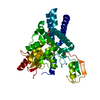
Mass: 65.409 Da / Num. of mol.: 2 / Source method: obtained synthetically / Formula: Zn
#3: Chemical ChemComp-Z3N / 2-{3-[(1s,3R)-3-methyl-1-(4-methyl-4H-1,2,4-triazol-3-yl)cyclobutyl]phenyl}-6-{[(3S)-3-methylpiperidin-1-yl]methyl}-4-(trifluoromethyl)-2,3-dihydro-1H-isoindol-1-one


Mass: 537.619 Da / Num. of mol.: 1 / Source method: obtained synthetically / Formula: C30H34F3N5O / Feature type: SUBJECT OF INVESTIGATION
#4: Chemical ChemComp-EDO / 1,2-ETHANEDIOL / ETHYLENE GLYCOL


Mass: 62.068 Da / Num. of mol.: 1 / Source method: obtained synthetically / Formula: C2H6O2
#5: Chemical ChemComp-SO4 / SULFATE ION


Mass: 96.063 Da / Num. of mol.: 1 / Source method: obtained synthetically / Formula: SO4
#6: Chemical ChemComp-UNX / UNKNOWN ATOM OR ION


Num. of mol.: 1 / Source method: obtained synthetically
#7: Water ChemComp-HOH / water


Mass: 18.015 Da / Num. of mol.: 142 / Source method: isolated from a natural source / Formula: H2O

-
Details

Has ligand of interestY

-
Experimental details

-
Experiment

ExperimentMethod: X-RAY DIFFRACTION / Number of used crystals: 1

-
Sample preparation

CrystalDensity Matthews: 2.13 Å3/Da / Density % sol: 42.25 %
Crystal growTemperature: 291 K / Method: vapor diffusion, sitting drop / pH: 6.5
Details: 0.1 M Imidazole, 0.1 M MES monohydrate pH 6.5 0.09 M Sodium nitrate, 0.09 M Sodium phosphate dibasic, 0.09M Ammonium sulfate 12.5% v/v MPD; 12.5% PEG1000; 12.5% w/v PEG 3350

-
Data collection

DiffractionMean temperature: 100 K / Serial crystal experiment: N
Diffraction sourceSource: SYNCHROTRON / Site: APS / Beamline: 24-ID-E / Wavelength: 0.97918 Å
DetectorType: DECTRIS EIGER2 X 16M / Detector: PIXEL / Date: Feb 11, 2023
RadiationProtocol: SINGLE WAVELENGTH / Monochromatic (M) / Laue (L): M / Scattering type: x-ray
Radiation wavelengthWavelength: 0.97918 Å / Relative weight: 1
ReflectionResolution: 1.81→50 Å / Num. obs: 35624 / % possible obs: 98.6 % / Redundancy: 10.9 % / CC1/2: 0.997 / CC star: 0.999 / Rmerge(I) obs: 0.064 / Χ2: 0.957 / Net I/σ(I): 36
Reflection shellResolution: 1.81→1.84 Å / Redundancy: 9.9 % / Rmerge(I) obs: 0.94 / Num. unique obs: 1755 / CC1/2: 0.827 / CC star: 0.951 / Χ2: 0.638 / % possible all: 98.3

-
Processing

Software
NameVersionClassification
HKL-3000data scaling
REFMACREFMAC 5.8.0258refinement
HKL-3000data reduction
PHASERphasing
RefinementMethod to determine structure: MOLECULAR REPLACEMENT / Resolution: 1.81→43.68 Å / Cor.coef. Fo:Fc: 0.961 / Cor.coef. Fo:Fc free: 0.943 / SU B: 3.58 / SU ML: 0.107 / Cross valid method: THROUGHOUT / ESU R: 0.142 / ESU R Free: 0.131 / Stereochemistry target values: MAXIMUM LIKELIHOOD / Details: HYDROGENS HAVE BEEN ADDED IN THE RIDING POSITIONS
RfactorNum. reflection% reflectionSelection details
Rfree0.22962 1795 5 %RANDOM
Rwork0.19491 ---
obs0.19669 33780 98.34 %-
Solvent computationIon probe radii: 0.8 Å / Shrinkage radii: 0.8 Å / VDW probe radii: 1.2 Å / Solvent model: MASK
Displacement parametersBiso mean: 32.702 Å2
Baniso -1Baniso -2Baniso -3
1--1.2 Å20 Å20 Å2
2--2.69 Å2-0 Å2
3----1.49 Å2
Refinement stepCycle: 1 / Resolution: 1.81→43.68 Å
ProteinNucleic acidLigandSolventTotal
Num. atoms2966 0 51 142 3159
Refine LS restraints
Refine-IDTypeDev idealDev ideal targetNumber
X-RAY DIFFRACTIONr_bond_refined_d0.0090.0133183
X-RAY DIFFRACTIONr_bond_other_d0.0010.0172851
X-RAY DIFFRACTIONr_angle_refined_deg1.4511.6414335
X-RAY DIFFRACTIONr_angle_other_deg1.3341.596594
X-RAY DIFFRACTIONr_dihedral_angle_1_deg6.4715392
X-RAY DIFFRACTIONr_dihedral_angle_2_deg33.06422.436156
X-RAY DIFFRACTIONr_dihedral_angle_3_deg13.88215512
X-RAY DIFFRACTIONr_dihedral_angle_4_deg18.0811516
X-RAY DIFFRACTIONr_chiral_restr0.0730.2412
X-RAY DIFFRACTIONr_gen_planes_refined0.0090.023636
X-RAY DIFFRACTIONr_gen_planes_other0.0020.02702
X-RAY DIFFRACTIONr_nbd_refined
X-RAY DIFFRACTIONr_nbd_other
X-RAY DIFFRACTIONr_nbtor_refined
X-RAY DIFFRACTIONr_nbtor_other
X-RAY DIFFRACTIONr_xyhbond_nbd_refined
X-RAY DIFFRACTIONr_xyhbond_nbd_other
X-RAY DIFFRACTIONr_metal_ion_refined
X-RAY DIFFRACTIONr_metal_ion_other
X-RAY DIFFRACTIONr_symmetry_vdw_refined
X-RAY DIFFRACTIONr_symmetry_vdw_other
X-RAY DIFFRACTIONr_symmetry_hbond_refined
X-RAY DIFFRACTIONr_symmetry_hbond_other
X-RAY DIFFRACTIONr_symmetry_metal_ion_refined
X-RAY DIFFRACTIONr_symmetry_metal_ion_other
X-RAY DIFFRACTIONr_mcbond_it3.0533.4541547
X-RAY DIFFRACTIONr_mcbond_other3.0393.4521546
X-RAY DIFFRACTIONr_mcangle_it4.1475.1631946
X-RAY DIFFRACTIONr_mcangle_other4.1485.1641947
X-RAY DIFFRACTIONr_scbond_it3.353.6721636
X-RAY DIFFRACTIONr_scbond_other3.3293.6691633
X-RAY DIFFRACTIONr_scangle_it
X-RAY DIFFRACTIONr_scangle_other4.9735.392382
X-RAY DIFFRACTIONr_long_range_B_refined6.61540.053630
X-RAY DIFFRACTIONr_long_range_B_other6.62139.9873610
X-RAY DIFFRACTIONr_rigid_bond_restr
X-RAY DIFFRACTIONr_sphericity_free
X-RAY DIFFRACTIONr_sphericity_bonded
LS refinement shellResolution: 1.81→1.853 Å
RfactorNum. reflection% reflection
Rfree0.265 130 -
Rwork0.273 2358 -
obs--94.46 %

+
About Yorodumi

-
News

-
Feb 9, 2022. New format data for meta-information of EMDB entries

New format data for meta-information of EMDB entries

  • Version 3 of the EMDB header file is now the official format.
  • The previous official version 1.9 will be removed from the archive.

Related info.:EMDB header

External links:wwPDB to switch to version 3 of the EMDB data model

-
Aug 12, 2020. Covid-19 info

Covid-19 info

URL: https://pdbjlvh1.pdbj.org/emnavi/covid19.php

New page: Covid-19 featured information page in EM Navigator.

Related info.:Covid-19 info / Mar 5, 2020. Novel coronavirus structure data

+
Mar 5, 2020. Novel coronavirus structure data

Novel coronavirus structure data

Related info.:Yorodumi Speices / Aug 12, 2020. Covid-19 info

External links:COVID-19 featured content - PDBj / Molecule of the Month (242):Coronavirus Proteases

+
Jan 31, 2019. EMDB accession codes are about to change! (news from PDBe EMDB page)

EMDB accession codes are about to change! (news from PDBe EMDB page)

  • The allocation of 4 digits for EMDB accession codes will soon come to an end. Whilst these codes will remain in use, new EMDB accession codes will include an additional digit and will expand incrementally as the available range of codes is exhausted. The current 4-digit format prefixed with “EMD-” (i.e. EMD-XXXX) will advance to a 5-digit format (i.e. EMD-XXXXX), and so on. It is currently estimated that the 4-digit codes will be depleted around Spring 2019, at which point the 5-digit format will come into force.
  • The EM Navigator/Yorodumi systems omit the EMD- prefix.

Related info.:Q: What is EMD? / ID/Accession-code notation in Yorodumi/EM Navigator

External links:EMDB Accession Codes are Changing Soon! / Contact to PDBj

+
Jul 12, 2017. Major update of PDB

Major update of PDB

  • wwPDB released updated PDB data conforming to the new PDBx/mmCIF dictionary.
  • This is a major update changing the version number from 4 to 5, and with Remediation, in which all the entries are updated.
  • In this update, many items about electron microscopy experimental information are reorganized (e.g. em_software).
  • Now, EM Navigator and Yorodumi are based on the updated data.

External links:wwPDB Remediation / Enriched Model Files Conforming to OneDep Data Standards Now Available in the PDB FTP Archive

-
Yorodumi

Thousand views of thousand structures

  • Yorodumi is a browser for structure data from EMDB, PDB, SASBDB, etc.
  • This page is also the successor to EM Navigator detail page, and also detail information page/front-end page for Omokage search.
  • The word "yorodu" (or yorozu) is an old Japanese word meaning "ten thousand". "mi" (miru) is to see.

Related info.:EMDB / PDB / SASBDB / Comparison of 3 databanks / Yorodumi Search / Aug 31, 2016. New EM Navigator & Yorodumi / Yorodumi Papers / Jmol/JSmol / Function and homology information / Changes in new EM Navigator and Yorodumi

Read more