 Movie
Movie Controller

[English] 日本語
 Yorodumi
- PDB-8fa0: Crystal structure of clade A/E 93TH057 HIV-1 gp120 core in comple... -
+ Open data

- Basic information
| Entry | Database: PDB / ID: 8fa0 | ||||||
|---|---|---|---|---|---|---|---|
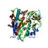
| Title | Crystal structure of clade A/E 93TH057 HIV-1 gp120 core in complex with NBD-14208, an HIV-1 gp120 antagonist | ||||||
 Components Components | HIV-1 gp120 core | ||||||
 Keywords Keywords | VIRAL PROTEIN / HIV-1 / NBD-14208 / small molecules / CD4-gp120 binding inhibitor / antagonist | ||||||
| Function / homology | HIV Envelope Protein Gp120; Chain G / Human immunodeficiency virus 1, Gp160, envelope glycoprotein / Gp120 core superfamily / Envelope glycoprotein GP120 / Human immunodeficiency virus 1, envelope glycoprotein Gp120 / Beta Complex / Mainly Beta / Chem-XKR / HIV-1 gp120 core Function and homology information Function and homology information | ||||||
| Biological species |  Homo sapiens (human) Homo sapiens (human) | ||||||
| Method |  X-RAY DIFFRACTION / SYNCHROTRON / MOLECULAR REPLACEMENT / Resolution: 2.09 Å X-RAY DIFFRACTION / SYNCHROTRON / MOLECULAR REPLACEMENT / Resolution: 2.09 Å | ||||||
 Authors Authors | Kwon, Y.D. / Kwong, P.D. | ||||||
| Funding support |  United States, 1items United States, 1items
| ||||||
 Citation Citation | Journal: Int J Mol Sci / Year: 2022 Title: Antiviral Activity and Crystal Structures of HIV-1 gp120 Antagonists. Authors: Curreli, F. / Kwon, Y.D. / Nicolau, I. / Burgos, G. / Altieri, A. / Kurkin, A.V. / Verardi, R. / Kwong, P.D. / Debnath, A.K. | ||||||
| History |
|
- Structure visualization
| Structure viewer | Molecule: MolmilJmol/JSmol |
|---|
- Downloads & links
-Download
| PDBx/mmCIF format | 8fa0.cif.gz | 157.5 KB | Display | PDBx/mmCIF format |
|---|---|---|---|---|
| PDB format | pdb8fa0.ent.gz | 122.2 KB | Display | PDB format |
| PDBx/mmJSON format | 8fa0.json.gz | Tree view | PDBx/mmJSON format | |
| Others |  Other downloads Other downloads |
-Validation report
| Arichive directory | https://data.pdbj.org/pub/pdb/validation_reports/fa/8fa0ftp://data.pdbj.org/pub/pdb/validation_reports/fa/8fa0 | HTTPS FTP |
|---|
-Related structure data
-Links
 PDBj
PDBj


- Assembly
| Deposited unit | 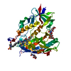
| ||||||||
|---|---|---|---|---|---|---|---|---|---|
| 1 |
| ||||||||
| Unit cell |
|
-Components
| #1: Protein | Mass: 39160.367 Da / Num. of mol.: 1 / Mutation: V1/V2, V3 deletion, H375S Source method: isolated from a genetically manipulated source Source: (gene. exp.) Homo sapiens (human) / Strain: clade A/E 93TH057 / Gene: HIV-1 Env / Plasmid: pVRC8400 / Cell (production host): 293F / Production host: Homo sapiens (human) / References: UniProt: A0A0M3KKW8 | ||||||||||
|---|---|---|---|---|---|---|---|---|---|---|---|
| #2: Sugar | ChemComp-NAG /   Type: D-saccharide, beta linking / Mass: 221.208 Da / Num. of mol.: 8 Type: D-saccharide, beta linking / Mass: 221.208 Da / Num. of mol.: 8Source method: isolated from a genetically manipulated source Formula: C8H15NO6 #3: Chemical |   Mass: 442.867 Da / Num. of mol.: 2 / Source method: obtained synthetically / Formula: C18H17ClF2N4O3S / Feature type: SUBJECT OF INVESTIGATION Mass: 442.867 Da / Num. of mol.: 2 / Source method: obtained synthetically / Formula: C18H17ClF2N4O3S / Feature type: SUBJECT OF INVESTIGATION#4: Chemical | ChemComp-EPE / |   Mass: 238.305 Da / Num. of mol.: 1 / Source method: obtained synthetically / Formula: C8H18N2O4S / Comment: pH buffer*YM Mass: 238.305 Da / Num. of mol.: 1 / Source method: obtained synthetically / Formula: C8H18N2O4S / Comment: pH buffer*YM#5: Water | ChemComp-HOH / |  Mass: 18.015 Da / Num. of mol.: 49 / Source method: isolated from a natural source / Formula: H2O Mass: 18.015 Da / Num. of mol.: 49 / Source method: isolated from a natural source / Formula: H2OHas ligand of interest | Y | Has protein modification | Y | |
-Experimental details
-Experiment
| Experiment | Method: X-RAY DIFFRACTION / Number of used crystals: 1 |
|---|
- Sample preparation
| Crystal | Density Matthews: 2.36 Å3/Da / Density % sol: 47.98 % |
|---|---|
| Crystal grow | Temperature: 293 K / Method: vapor diffusion, hanging drop / pH: 7.4 / Details: 12-14% PEG3350, 5% isopropanol, 0.1M HEPES, 7.4 |
-Data collection
| Diffraction | Mean temperature: 100 K / Serial crystal experiment: N | |||||||||||||||||||||||||||||||||||||||||||||||||||||||||||||||||||||||||||||||||||||||||||||||||||||||||||||||||||||||||||||||||||||||||||||||||||
|---|---|---|---|---|---|---|---|---|---|---|---|---|---|---|---|---|---|---|---|---|---|---|---|---|---|---|---|---|---|---|---|---|---|---|---|---|---|---|---|---|---|---|---|---|---|---|---|---|---|---|---|---|---|---|---|---|---|---|---|---|---|---|---|---|---|---|---|---|---|---|---|---|---|---|---|---|---|---|---|---|---|---|---|---|---|---|---|---|---|---|---|---|---|---|---|---|---|---|---|---|---|---|---|---|---|---|---|---|---|---|---|---|---|---|---|---|---|---|---|---|---|---|---|---|---|---|---|---|---|---|---|---|---|---|---|---|---|---|---|---|---|---|---|---|---|---|---|---|
| Diffraction source | Source: SYNCHROTRON / Site: APS / Beamline: 22-ID / Wavelength: 1 Å | |||||||||||||||||||||||||||||||||||||||||||||||||||||||||||||||||||||||||||||||||||||||||||||||||||||||||||||||||||||||||||||||||||||||||||||||||||
| Detector | Type: DECTRIS EIGER X 16M / Detector: PIXEL / Date: Nov 17, 2017 | |||||||||||||||||||||||||||||||||||||||||||||||||||||||||||||||||||||||||||||||||||||||||||||||||||||||||||||||||||||||||||||||||||||||||||||||||||
| Radiation | Protocol: SINGLE WAVELENGTH / Monochromatic (M) / Laue (L): M / Scattering type: x-ray | |||||||||||||||||||||||||||||||||||||||||||||||||||||||||||||||||||||||||||||||||||||||||||||||||||||||||||||||||||||||||||||||||||||||||||||||||||
| Radiation wavelength | Wavelength: 1 Å / Relative weight: 1 | |||||||||||||||||||||||||||||||||||||||||||||||||||||||||||||||||||||||||||||||||||||||||||||||||||||||||||||||||||||||||||||||||||||||||||||||||||
| Reflection | Resolution: 2.09→50 Å / Num. obs: 39369 / % possible obs: 93.7 % / Redundancy: 3.5 % / Rmerge(I) obs: 0.161 / Χ2: 0.102 / Net I/σ(I): 7 | |||||||||||||||||||||||||||||||||||||||||||||||||||||||||||||||||||||||||||||||||||||||||||||||||||||||||||||||||||||||||||||||||||||||||||||||||||
| Reflection shell |
|
- Processing
| Software |
| |||||||||||||||||||||||||||||||||||||||||||||||||||||||||||||||||||||||||||||||||||||||||||||||||||||||||||||||||||||||||||||
|---|---|---|---|---|---|---|---|---|---|---|---|---|---|---|---|---|---|---|---|---|---|---|---|---|---|---|---|---|---|---|---|---|---|---|---|---|---|---|---|---|---|---|---|---|---|---|---|---|---|---|---|---|---|---|---|---|---|---|---|---|---|---|---|---|---|---|---|---|---|---|---|---|---|---|---|---|---|---|---|---|---|---|---|---|---|---|---|---|---|---|---|---|---|---|---|---|---|---|---|---|---|---|---|---|---|---|---|---|---|---|---|---|---|---|---|---|---|---|---|---|---|---|---|---|---|---|
| Refinement | Method to determine structure: MOLECULAR REPLACEMENT / Resolution: 2.09→36.35 Å / SU ML: 0.35 / Cross valid method: FREE R-VALUE / σ(F): 1.34 / Phase error: 38 / Stereochemistry target values: ML
| |||||||||||||||||||||||||||||||||||||||||||||||||||||||||||||||||||||||||||||||||||||||||||||||||||||||||||||||||||||||||||||
| Solvent computation | Shrinkage radii: 0.9 Å / VDW probe radii: 1.11 Å / Solvent model: FLAT BULK SOLVENT MODEL | |||||||||||||||||||||||||||||||||||||||||||||||||||||||||||||||||||||||||||||||||||||||||||||||||||||||||||||||||||||||||||||
| Refinement step | Cycle: LAST / Resolution: 2.09→36.35 Å
| |||||||||||||||||||||||||||||||||||||||||||||||||||||||||||||||||||||||||||||||||||||||||||||||||||||||||||||||||||||||||||||
| Refine LS restraints |
| |||||||||||||||||||||||||||||||||||||||||||||||||||||||||||||||||||||||||||||||||||||||||||||||||||||||||||||||||||||||||||||
| LS refinement shell |
| |||||||||||||||||||||||||||||||||||||||||||||||||||||||||||||||||||||||||||||||||||||||||||||||||||||||||||||||||||||||||||||
| Refinement TLS params. | Method: refined / Refine-ID: X-RAY DIFFRACTION
| |||||||||||||||||||||||||||||||||||||||||||||||||||||||||||||||||||||||||||||||||||||||||||||||||||||||||||||||||||||||||||||
| Refinement TLS group |
|
