 Movie
Movie Controller

[English] 日本語
 Yorodumi
- PDB-8dnj: Crystal structure of human KRAS G12C covalently bound with AstraZ... -
+ Open data

- Basic information
| Entry | Database: PDB / ID: 8dnj | ||||||
|---|---|---|---|---|---|---|---|
| Title | Crystal structure of human KRAS G12C covalently bound with AstraZeneca WO2020/178282A1 compound 76 | ||||||
 Components Components | Isoform 2B of GTPase KRas | ||||||
 Keywords Keywords | SIGNALING PROTEIN / Inhibitor / GTPase | ||||||
| Function / homology | small monomeric GTPase / Ca2+ pathway / GUANOSINE-5'-DIPHOSPHATE / Chem-U4U / Isoform 2B of GTPase KRas Function and homology information Function and homology information | ||||||
| Biological species |  Homo sapiens (human) Homo sapiens (human) | ||||||
| Method |  X-RAY DIFFRACTION / SYNCHROTRON / MOLECULAR REPLACEMENT / Resolution: 1.81 Å X-RAY DIFFRACTION / SYNCHROTRON / MOLECULAR REPLACEMENT / Resolution: 1.81 Å | ||||||
 Authors Authors | Mohr, C. | ||||||
| Funding support | 1items
| ||||||
 Citation Citation | Journal: J.Comput.Aided Mol.Des. / Year: 2022 Title: Modeling receptor flexibility in the structure-based design of KRAS G12C inhibitors. Authors: Zhu, K. / Li, C. / Wu, K.Y. / Mohr, C. / Li, X. / Lanman, B. | ||||||
| History |
|
- Structure visualization
| Structure viewer | Molecule: MolmilJmol/JSmol |
|---|
- Downloads & links
-Download
| PDBx/mmCIF format | 8dnj.cif.gz | 124 KB | Display | PDBx/mmCIF format |
|---|---|---|---|---|
| PDB format | pdb8dnj.ent.gz | 93.5 KB | Display | PDB format |
| PDBx/mmJSON format | 8dnj.json.gz | Tree view | PDBx/mmJSON format | |
| Others |  Other downloads Other downloads |
-Validation report
| Summary document | 8dnj_validation.pdf.gz | 2.1 MB | Display | wwPDB validaton report |
|---|---|---|---|---|
| Full document | 8dnj_full_validation.pdf.gz | 2.1 MB | Display | |
| Data in XML | 8dnj_validation.xml.gz | 23.7 KB | Display | |
| Data in CIF | 8dnj_validation.cif.gz | 31.9 KB | Display | |
| Arichive directory | https://data.pdbj.org/pub/pdb/validation_reports/dn/8dnjftp://data.pdbj.org/pub/pdb/validation_reports/dn/8dnj | HTTPS FTP |
-Related structure data
| Related structure data |  8dniC  8dnkC  6oimS C: citing same article ( S: Starting model for refinement |
|---|---|
| Similar structure data |
-Links
 PDBj
PDBj


- Assembly
| Deposited unit | 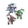
| ||||||||
|---|---|---|---|---|---|---|---|---|---|
| 1 | 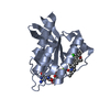
| ||||||||
| 2 | 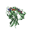
| ||||||||
| 3 | 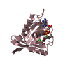
| ||||||||
| Unit cell |
|
-Components
| #1: Protein | Mass: 21133.867 Da / Num. of mol.: 3 / Mutation: C51S,C80L,C118S Source method: isolated from a genetically manipulated source Details: G12C variant / Source: (gene. exp.) Homo sapiens (human) / Gene: KRAS, KRAS2, RASK2 / Variant: VAR_006839 G12C / Production host:  #2: Chemical |   Mass: 24.305 Da / Num. of mol.: 3 / Source method: obtained synthetically / Formula: Mg Mass: 24.305 Da / Num. of mol.: 3 / Source method: obtained synthetically / Formula: Mg#3: Chemical |   Type: RNA linking / Mass: 443.201 Da / Num. of mol.: 3 / Source method: obtained synthetically / Formula: C10H15N5O11P2 / Comment: GDP, energy-carrying molecule*YM Type: RNA linking / Mass: 443.201 Da / Num. of mol.: 3 / Source method: obtained synthetically / Formula: C10H15N5O11P2 / Comment: GDP, energy-carrying molecule*YM#4: Chemical |   Mass: 428.884 Da / Num. of mol.: 3 / Source method: obtained synthetically / Formula: C23H22ClFN2O3 / Feature type: SUBJECT OF INVESTIGATION Mass: 428.884 Da / Num. of mol.: 3 / Source method: obtained synthetically / Formula: C23H22ClFN2O3 / Feature type: SUBJECT OF INVESTIGATION#5: Water | ChemComp-HOH / |  Mass: 18.015 Da / Num. of mol.: 199 / Source method: isolated from a natural source / Formula: H2O Mass: 18.015 Da / Num. of mol.: 199 / Source method: isolated from a natural source / Formula: H2OHas ligand of interest | Y | |
|---|
-Experimental details
-Experiment
| Experiment | Method: X-RAY DIFFRACTION / Number of used crystals: 1 |
|---|
- Sample preparation
| Crystal | Density Matthews: 1.91 Å3/Da / Density % sol: 35.58 % |
|---|---|
| Crystal grow | Temperature: 293 K / Method: vapor diffusion, sitting drop / pH: 6.5 Details: 0.001M MgCl2, 0.1 M MES pH 6.5, 30% PEG 4000, 10% Ethanol |
-Data collection
| Diffraction | Mean temperature: 100 K / Serial crystal experiment: N |
|---|---|
| Diffraction source | Source: SYNCHROTRON / Site: ALS  / Beamline: 5.0.2 / Wavelength: 0.99999 Å / Beamline: 5.0.2 / Wavelength: 0.99999 Å |
| Detector | Type: DECTRIS PILATUS3 6M / Detector: PIXEL / Date: Dec 18, 2020 |
| Radiation | Monochromator: Double-crystal, Si(111) / Protocol: SINGLE WAVELENGTH / Monochromatic (M) / Laue (L): M / Scattering type: x-ray |
| Radiation wavelength | Wavelength: 0.99999 Å / Relative weight: 1 |
| Reflection | Resolution: 1.81→112.11 Å / Num. obs: 43236 / % possible obs: 99.8 % / Redundancy: 6.3 % / CC1/2: 0.948 / Net I/σ(I): 4.8 |
| Reflection shell | Resolution: 1.81→1.84 Å / Redundancy: 5.3 % / Mean I/σ(I) obs: 1.1 / Num. unique obs: 1996 / CC1/2: 0.564 / % possible all: 95.2 |
- Processing
| Software |
| ||||||||||||||||||||||||||||||||||||||||||||||||||||||||||||
|---|---|---|---|---|---|---|---|---|---|---|---|---|---|---|---|---|---|---|---|---|---|---|---|---|---|---|---|---|---|---|---|---|---|---|---|---|---|---|---|---|---|---|---|---|---|---|---|---|---|---|---|---|---|---|---|---|---|---|---|---|---|
| Refinement | Method to determine structure: MOLECULAR REPLACEMENT Starting model: 6OIM Resolution: 1.81→30 Å / Cor.coef. Fo:Fc: 0.935 / Cor.coef. Fo:Fc free: 0.896 / SU B: 7.927 / SU ML: 0.214 / Cross valid method: THROUGHOUT / σ(F): 0 / ESU R: 0.193 / ESU R Free: 0.175 / Stereochemistry target values: MAXIMUM LIKELIHOOD Details: HYDROGENS HAVE BEEN ADDED IN THE RIDING POSITIONS U VALUES : REFINED INDIVIDUALLY
| ||||||||||||||||||||||||||||||||||||||||||||||||||||||||||||
| Solvent computation | Ion probe radii: 0.8 Å / Shrinkage radii: 0.8 Å / VDW probe radii: 1.2 Å / Solvent model: MASK | ||||||||||||||||||||||||||||||||||||||||||||||||||||||||||||
| Displacement parameters | Biso max: 70.31 Å2 / Biso mean: 25.834 Å2 / Biso min: 12.54 Å2
| ||||||||||||||||||||||||||||||||||||||||||||||||||||||||||||
| Refinement step | Cycle: final / Resolution: 1.81→30 Å
| ||||||||||||||||||||||||||||||||||||||||||||||||||||||||||||
| Refine LS restraints |
| ||||||||||||||||||||||||||||||||||||||||||||||||||||||||||||
| LS refinement shell | Resolution: 1.81→1.857 Å / Rfactor Rfree error: 0 / Total num. of bins used: 20
|
