 Movie
Movie Controller

[English] 日本語
 Yorodumi
- PDB-8dhn: The N-terminal domain of PA endonuclease from the influenza H1N1 ... -
+ Open data

- Basic information
| Entry | Database: PDB / ID: 8dhn | ||||||
|---|---|---|---|---|---|---|---|
| Title | The N-terminal domain of PA endonuclease from the influenza H1N1 viral polymerase in complex with 6-Bromo-3-hydroxy-2-(5-methyl-1,2,4-oxadiazol-3-yl)pyridin-4(1H)-one | ||||||
 Components Components | PA endonuclease | ||||||
 Keywords Keywords | VIRAL PROTEIN / HYDROLASE/INHIBITOR / Drug discovery / metal-binding pharmacophore / isosteres / influenza endonuclease / VIRAL PROTEIN-INHIBITOR complex / ANTIVIRAL PROTEIN / HYDROLASE-INHIBITOR complex | ||||||
| Function / homology | : / Chem-T6U Function and homology information Function and homology information | ||||||
| Biological species |   Influenza A virus Influenza A virus | ||||||
| Method |  X-RAY DIFFRACTION / MOLECULAR REPLACEMENT / Resolution: 2.4 Å X-RAY DIFFRACTION / MOLECULAR REPLACEMENT / Resolution: 2.4 Å | ||||||
 Authors Authors | Kohlbrand, A.J. / Stokes, R.W. / Karges, J. / Seo, H. / Sankaran, B. / Cohen, S.M. | ||||||
| Funding support |  United States, 1items United States, 1items
| ||||||
 Citation Citation | Journal: Acs Med.Chem.Lett. / Year: 2023 Title: Carboxylic Acid Isostere Derivatives of Hydroxypyridinones as Core Scaffolds for Influenza Endonuclease Inhibitors. Authors: Stokes, R.W. / Kohlbrand, A.J. / Seo, H. / Sankaran, B. / Karges, J. / Cohen, S.M. | ||||||
| History |
|
- Structure visualization
| Structure viewer | Molecule: MolmilJmol/JSmol |
|---|
- Downloads & links
-Download
| PDBx/mmCIF format | 8dhn.cif.gz | 105.4 KB | Display | PDBx/mmCIF format |
|---|---|---|---|---|
| PDB format | pdb8dhn.ent.gz | 69.2 KB | Display | PDB format |
| PDBx/mmJSON format | 8dhn.json.gz | Tree view | PDBx/mmJSON format | |
| Others |  Other downloads Other downloads |
-Validation report
| Arichive directory | https://data.pdbj.org/pub/pdb/validation_reports/dh/8dhnftp://data.pdbj.org/pub/pdb/validation_reports/dh/8dhn | HTTPS FTP |
|---|
-Related structure data
| Related structure data | 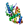 7v04C 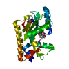 8ctfC 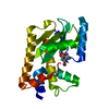 8dalC 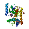 8ddbC 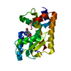 8ddeC 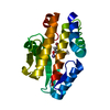 8djvC 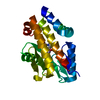 8djyC  6e6vS C: citing same article ( S: Starting model for refinement |
|---|---|
| Similar structure data |
-Links
 PDBj
PDBj- Assembly
| Deposited unit | 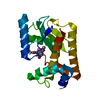
| ||||||||||||
|---|---|---|---|---|---|---|---|---|---|---|---|---|---|
| 1 |
| ||||||||||||
| Unit cell |
| ||||||||||||
| Components on special symmetry positions |
|
-Components
| #1: Protein | Mass: 22424.547 Da / Num. of mol.: 1 / Fragment: N-terminal domain Source method: isolated from a genetically manipulated source Source: (gene. exp.) Influenza A virus / Strain: A/California/04/2009(H1N1) / Production host:  | ||||
|---|---|---|---|---|---|
| #2: Chemical | ChemComp-T6U / (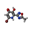  Mass: 272.056 Da / Num. of mol.: 1 / Source method: obtained synthetically / Formula: C8H6BrN3O3 / Feature type: SUBJECT OF INVESTIGATION Mass: 272.056 Da / Num. of mol.: 1 / Source method: obtained synthetically / Formula: C8H6BrN3O3 / Feature type: SUBJECT OF INVESTIGATION | ||||
| #3: Chemical |   Mass: 54.938 Da / Num. of mol.: 2 / Source method: obtained synthetically / Formula: Mn Mass: 54.938 Da / Num. of mol.: 2 / Source method: obtained synthetically / Formula: Mn#4: Water | ChemComp-HOH / |  Mass: 18.015 Da / Num. of mol.: 83 / Source method: isolated from a natural source / Formula: H2O Mass: 18.015 Da / Num. of mol.: 83 / Source method: isolated from a natural source / Formula: H2OHas ligand of interest | Y | |
-Experimental details
-Experiment
| Experiment | Method: X-RAY DIFFRACTION / Number of used crystals: 1 |
|---|
- Sample preparation
| Crystal | Density Matthews: 2.21 Å3/Da / Density % sol: 44.33 % |
|---|---|
| Crystal grow | Temperature: 293 K / Method: vapor diffusion, hanging drop Details: 32% PEG4000, 100 mM Tris, pH 8.35, 200-220 mM sodium acetate |
-Data collection
| Diffraction | Mean temperature: 100 K / Serial crystal experiment: N |
|---|---|
| Diffraction source | Source: ROTATING ANODE / Type: BRUKER AXS MICROSTAR / Wavelength: 1.54 Å |
| Detector | Type: APEX II CCD / Detector: CCD / Date: Jun 15, 2021 |
| Radiation | Protocol: SINGLE WAVELENGTH / Monochromatic (M) / Laue (L): M / Scattering type: x-ray |
| Radiation wavelength | Wavelength: 1.54 Å / Relative weight: 1 |
| Reflection | Resolution: 2.4→65.01 Å / Num. obs: 15004 / % possible obs: 99.9 % / Redundancy: 29 % / Biso Wilson estimate: 47.41 Å2 / CC1/2: 1 / Rmerge(I) obs: 0.1 / Rrim(I) all: 0.102 / Net I/σ(I): 20 |
| Reflection shell | Resolution: 2.4→2.48 Å / Rmerge(I) obs: 1.439 / Mean I/σ(I) obs: 2 / Num. unique obs: 806 / CC1/2: 0.84 / Rpim(I) all: 0.33 |
- Processing
| Software |
| ||||||||||||||||||||||||||||||||||||||||||||||||||||||||||||||||||||||||||||||||||||||||||||||||||||
|---|---|---|---|---|---|---|---|---|---|---|---|---|---|---|---|---|---|---|---|---|---|---|---|---|---|---|---|---|---|---|---|---|---|---|---|---|---|---|---|---|---|---|---|---|---|---|---|---|---|---|---|---|---|---|---|---|---|---|---|---|---|---|---|---|---|---|---|---|---|---|---|---|---|---|---|---|---|---|---|---|---|---|---|---|---|---|---|---|---|---|---|---|---|---|---|---|---|---|---|---|---|
| Refinement | Method to determine structure: MOLECULAR REPLACEMENT Starting model: 6E6V Resolution: 2.4→64.98 Å / SU ML: 0.2803 / Cross valid method: FREE R-VALUE / σ(F): 1.33 / Phase error: 29.7585 Stereochemistry target values: GeoStd + Monomer Library + CDL v1.2
| ||||||||||||||||||||||||||||||||||||||||||||||||||||||||||||||||||||||||||||||||||||||||||||||||||||
| Solvent computation | Shrinkage radii: 0.9 Å / VDW probe radii: 1.11 Å / Solvent model: FLAT BULK SOLVENT MODEL | ||||||||||||||||||||||||||||||||||||||||||||||||||||||||||||||||||||||||||||||||||||||||||||||||||||
| Displacement parameters | Biso mean: 54.05 Å2 | ||||||||||||||||||||||||||||||||||||||||||||||||||||||||||||||||||||||||||||||||||||||||||||||||||||
| Refinement step | Cycle: LAST / Resolution: 2.4→64.98 Å
| ||||||||||||||||||||||||||||||||||||||||||||||||||||||||||||||||||||||||||||||||||||||||||||||||||||
| Refine LS restraints |
| ||||||||||||||||||||||||||||||||||||||||||||||||||||||||||||||||||||||||||||||||||||||||||||||||||||
| LS refinement shell |
| ||||||||||||||||||||||||||||||||||||||||||||||||||||||||||||||||||||||||||||||||||||||||||||||||||||
| Refinement TLS params. | Method: refined / Refine-ID: X-RAY DIFFRACTION
| ||||||||||||||||||||||||||||||||||||||||||||||||||||||||||||||||||||||||||||||||||||||||||||||||||||
| Refinement TLS group | Refine-ID: X-RAY DIFFRACTION / Auth asym-ID: A / Label asym-ID: B
|
