 Movie
Movie Controller

[English] 日本語
 Yorodumi
- PDB-8aj3: X-ray structure of lysozyme obtained upon reaction with [VIVO(mal... -
+ Open data

- Basic information
| Entry | Database: PDB / ID: 8aj3 | ||||||
|---|---|---|---|---|---|---|---|
| Title | X-ray structure of lysozyme obtained upon reaction with [VIVO(malt)2] (Structure A) | ||||||
 Components Components | Lysozyme | ||||||
 Keywords Keywords | HYDROLASE / metallodrug / vanadium / oxidovanadium / covalent binding / non-covalent binding / protein-metal interaction | ||||||
| Function / homology |  Function and homology information Function and homology informationLactose synthesis / Antimicrobial peptides / Neutrophil degranulation / beta-N-acetylglucosaminidase activity / cell wall macromolecule catabolic process / lysozyme / lysozyme activity / defense response to Gram-negative bacterium / killing of cells of another organism / defense response to Gram-positive bacterium ...Lactose synthesis / Antimicrobial peptides / Neutrophil degranulation / beta-N-acetylglucosaminidase activity / cell wall macromolecule catabolic process / lysozyme / lysozyme activity / defense response to Gram-negative bacterium / killing of cells of another organism / defense response to Gram-positive bacterium / defense response to bacterium / endoplasmic reticulum / extracellular space / identical protein binding / cytoplasm Similarity search - Function | ||||||
| Biological species |  | ||||||
| Method |  X-RAY DIFFRACTION / SYNCHROTRON / MOLECULAR REPLACEMENT / Resolution: 1.13 Å X-RAY DIFFRACTION / SYNCHROTRON / MOLECULAR REPLACEMENT / Resolution: 1.13 Å | ||||||
 Authors Authors | Paolillo, M. / Merlino, A. / Ferraro, G. | ||||||
| Funding support | 1items
| ||||||
 Citation Citation | Journal: Inorg.Chem. / Year: 2022 Title: Multiple and Variable Binding of Pharmacologically Active Bis(maltolato)oxidovanadium(IV) to Lysozyme. Authors: Ferraro, G. / Paolillo, M. / Sciortino, G. / Garribba, E. / Merlino, A. | ||||||
| History |
|
- Structure visualization
| Structure viewer | Molecule: MolmilJmol/JSmol |
|---|
- Downloads & links
-Download
| PDBx/mmCIF format | 8aj3.cif.gz | 75 KB | Display | PDBx/mmCIF format |
|---|---|---|---|---|
| PDB format | pdb8aj3.ent.gz | 52.1 KB | Display | PDB format |
| PDBx/mmJSON format | 8aj3.json.gz | Tree view | PDBx/mmJSON format | |
| Others |  Other downloads Other downloads |
-Validation report
| Summary document | 8aj3_validation.pdf.gz | 1.6 MB | Display | wwPDB validaton report |
|---|---|---|---|---|
| Full document | 8aj3_full_validation.pdf.gz | 1.6 MB | Display | |
| Data in XML | 8aj3_validation.xml.gz | 9.5 KB | Display | |
| Data in CIF | 8aj3_validation.cif.gz | 13.5 KB | Display | |
| Arichive directory | https://data.pdbj.org/pub/pdb/validation_reports/aj/8aj3ftp://data.pdbj.org/pub/pdb/validation_reports/aj/8aj3 | HTTPS FTP |
-Related structure data
| Related structure data | 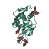 8aj4C 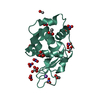 8aj5C 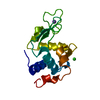 193lS S: Starting model for refinement C: citing same article ( |
|---|---|
| Similar structure data |
-Links
 PDBj
PDBj


- Assembly
| Deposited unit | 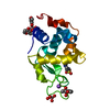
| ||||||||||||
|---|---|---|---|---|---|---|---|---|---|---|---|---|---|
| 1 |
| ||||||||||||
| Unit cell |
| ||||||||||||
| Components on special symmetry positions |
|
-Components
-Protein , 1 types, 1 molecules A
| #1: Protein | Mass: 14331.160 Da / Num. of mol.: 1 / Source method: isolated from a natural source / Source: (natural) |
|---|
-Non-polymers , 6 types, 163 molecules 


| #2: Chemical | ChemComp-NA /  Mass: 22.990 Da / Num. of mol.: 1 / Source method: obtained synthetically / Formula: Na Mass: 22.990 Da / Num. of mol.: 1 / Source method: obtained synthetically / Formula: Na |
|---|---|
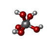
| #3: Chemical | ChemComp-H1W /  Mass: 135.978 Da / Num. of mol.: 1 / Source method: obtained synthetically / Formula: H5O5V / Feature type: SUBJECT OF INVESTIGATION Mass: 135.978 Da / Num. of mol.: 1 / Source method: obtained synthetically / Formula: H5O5V / Feature type: SUBJECT OF INVESTIGATION |
| #4: Chemical | ChemComp-EPE /  Mass: 238.305 Da / Num. of mol.: 1 / Source method: obtained synthetically / Formula: C8H18N2O4S / Comment: pH buffer*YM Mass: 238.305 Da / Num. of mol.: 1 / Source method: obtained synthetically / Formula: C8H18N2O4S / Comment: pH buffer*YM |
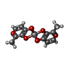
| #5: Chemical | ChemComp-MKO /  Mass: 333.145 Da / Num. of mol.: 1 / Source method: obtained synthetically / Formula: C12H10O8V / Feature type: SUBJECT OF INVESTIGATION Mass: 333.145 Da / Num. of mol.: 1 / Source method: obtained synthetically / Formula: C12H10O8V / Feature type: SUBJECT OF INVESTIGATION |
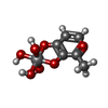
| #6: Chemical | ChemComp-MNW /  Mass: 244.073 Da / Num. of mol.: 1 / Source method: obtained synthetically / Formula: C6H9O7V / Feature type: SUBJECT OF INVESTIGATION Mass: 244.073 Da / Num. of mol.: 1 / Source method: obtained synthetically / Formula: C6H9O7V / Feature type: SUBJECT OF INVESTIGATION |
| #7: Water | ChemComp-HOH / Mass: 18.015 Da / Num. of mol.: 158 / Source method: isolated from a natural source / Formula: H2O |
-Details
| Has ligand of interest | Y |
|---|
-Experimental details
-Experiment
| Experiment | Method: X-RAY DIFFRACTION / Number of used crystals: 1 |
|---|
- Sample preparation
| Crystal | Density Matthews: 1.96 Å3/Da / Density % sol: 37.31 % |
|---|---|
| Crystal grow | Temperature: 293 K / Method: vapor diffusion, hanging drop / pH: 4 / Details: 2.0 M sodium formate and 0.1 M Hepes, pH 7.5 |
-Data collection
| Diffraction | Mean temperature: 100 K / Serial crystal experiment: N |
|---|---|
| Diffraction source | Source: SYNCHROTRON / Site: ELETTRA  / Beamline: 11.2C / Wavelength: 1 Å / Beamline: 11.2C / Wavelength: 1 Å |
| Detector | Type: DECTRIS PILATUS 6M / Detector: PIXEL / Date: Jul 28, 2021 |
| Radiation | Protocol: SINGLE WAVELENGTH / Monochromatic (M) / Laue (L): M / Scattering type: x-ray |
| Radiation wavelength | Wavelength: 1 Å / Relative weight: 1 |
| Reflection | Resolution: 1.13→54.37 Å / Num. obs: 42289 / % possible obs: 97.3 % / Redundancy: 16.1 % / CC1/2: 0.999 / Rmerge(I) obs: 0.06 / Net I/σ(I): 28.4 |
| Reflection shell | Resolution: 1.13→1.15 Å / Redundancy: 5 % / Rmerge(I) obs: 0.339 / Mean I/σ(I) obs: 3.4 / Num. unique obs: 1866 / CC1/2: 0.926 / % possible all: 87.9 |
- Processing
| Software |
| |||||||||||||||||||||||||||||||||||||||||||||||||||||||||||||||||||||||||||||||||||||||||||||||||||||||||||||||||||||||||||||||||||||||||||||||||||||||||||||||||||||||||||||||||||||||||||||||||||||||||||||||||||||||||||||||||||||||
|---|---|---|---|---|---|---|---|---|---|---|---|---|---|---|---|---|---|---|---|---|---|---|---|---|---|---|---|---|---|---|---|---|---|---|---|---|---|---|---|---|---|---|---|---|---|---|---|---|---|---|---|---|---|---|---|---|---|---|---|---|---|---|---|---|---|---|---|---|---|---|---|---|---|---|---|---|---|---|---|---|---|---|---|---|---|---|---|---|---|---|---|---|---|---|---|---|---|---|---|---|---|---|---|---|---|---|---|---|---|---|---|---|---|---|---|---|---|---|---|---|---|---|---|---|---|---|---|---|---|---|---|---|---|---|---|---|---|---|---|---|---|---|---|---|---|---|---|---|---|---|---|---|---|---|---|---|---|---|---|---|---|---|---|---|---|---|---|---|---|---|---|---|---|---|---|---|---|---|---|---|---|---|---|---|---|---|---|---|---|---|---|---|---|---|---|---|---|---|---|---|---|---|---|---|---|---|---|---|---|---|---|---|---|---|---|---|---|---|---|---|---|---|---|---|---|---|---|---|---|---|---|---|
| Refinement | Method to determine structure: MOLECULAR REPLACEMENT Starting model: 193L Resolution: 1.13→54.369 Å / Cor.coef. Fo:Fc: 0.953 / Cor.coef. Fo:Fc free: 0.933 / SU B: 2.438 / SU ML: 0.049 / Cross valid method: FREE R-VALUE / ESU R: 0.048 / ESU R Free: 0.051 Details: Hydrogens have been used if present in the input file
| |||||||||||||||||||||||||||||||||||||||||||||||||||||||||||||||||||||||||||||||||||||||||||||||||||||||||||||||||||||||||||||||||||||||||||||||||||||||||||||||||||||||||||||||||||||||||||||||||||||||||||||||||||||||||||||||||||||||
| Solvent computation | Ion probe radii: 0.8 Å / Shrinkage radii: 0.8 Å / VDW probe radii: 1.2 Å / Solvent model: MASK BULK SOLVENT | |||||||||||||||||||||||||||||||||||||||||||||||||||||||||||||||||||||||||||||||||||||||||||||||||||||||||||||||||||||||||||||||||||||||||||||||||||||||||||||||||||||||||||||||||||||||||||||||||||||||||||||||||||||||||||||||||||||||
| Displacement parameters | Biso mean: 16.146 Å2
| |||||||||||||||||||||||||||||||||||||||||||||||||||||||||||||||||||||||||||||||||||||||||||||||||||||||||||||||||||||||||||||||||||||||||||||||||||||||||||||||||||||||||||||||||||||||||||||||||||||||||||||||||||||||||||||||||||||||
| Refinement step | Cycle: LAST / Resolution: 1.13→54.369 Å
| |||||||||||||||||||||||||||||||||||||||||||||||||||||||||||||||||||||||||||||||||||||||||||||||||||||||||||||||||||||||||||||||||||||||||||||||||||||||||||||||||||||||||||||||||||||||||||||||||||||||||||||||||||||||||||||||||||||||
| Refine LS restraints |
| |||||||||||||||||||||||||||||||||||||||||||||||||||||||||||||||||||||||||||||||||||||||||||||||||||||||||||||||||||||||||||||||||||||||||||||||||||||||||||||||||||||||||||||||||||||||||||||||||||||||||||||||||||||||||||||||||||||||
| LS refinement shell | Refine-ID: X-RAY DIFFRACTION / Total num. of bins used: 20
|
