 Movie
Movie Controller


+ Open data

- Basic information
| Entry | Database: EMDB / ID: EMD-8410 | |||||||||
|---|---|---|---|---|---|---|---|---|---|---|
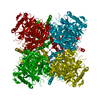
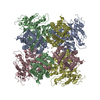
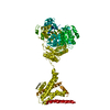
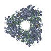
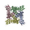
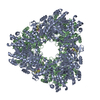
| Title | Ca2+ bound aplysia Slo1 | |||||||||
 Map data Map data | Mg and Ca-bound aplysia Slo1 | |||||||||
 Sample Sample |
| |||||||||
 Keywords Keywords | ion channel / K+ channel / Ca2+ bound / high conductance / MEMBRANE PROTEIN | |||||||||
| Function / homology |  Function and homology information Function and homology informationlarge conductance calcium-activated potassium channel activity / monoatomic ion channel complex / modulation of chemical synaptic transmission / postsynaptic membrane / response to xenobiotic stimulus / metal ion binding Similarity search - Function | |||||||||
| Biological species |  | |||||||||
| Method | single particle reconstruction / cryo EM / Resolution: 3.5 Å | |||||||||
 Authors Authors | MacKinnon R / Tao X | |||||||||
| Funding support |  United States, 2 items United States, 2 items
| |||||||||
 Citation Citation | Journal: Nature / Year: 2017 Title: Cryo-EM structure of the open high-conductance Ca-activated K channel. Authors: Xiao Tao / Richard K Hite / Roderick MacKinnon / Abstract: The Ca-activated K channel, Slo1, has an unusually large conductance and contains a voltage sensor and multiple chemical sensors. Dual activation by membrane voltage and Ca renders Slo1 central to ...The Ca-activated K channel, Slo1, has an unusually large conductance and contains a voltage sensor and multiple chemical sensors. Dual activation by membrane voltage and Ca renders Slo1 central to processes that couple electrical signalling to Ca-mediated events such as muscle contraction and neuronal excitability. Here we present the cryo-electron microscopy structure of a full-length Slo1 channel from Aplysia californica in the presence of Ca and Mg at a resolution of 3.5 Å. The channel adopts an open conformation. Its voltage-sensor domain adopts a non-domain-swapped attachment to the pore and contacts the cytoplasmic Ca-binding domain from a neighbouring subunit. Unique structural features of the Slo1 voltage sensor suggest that it undergoes different conformational changes than other known voltage sensors. The structure reveals the molecular details of three distinct divalent cation-binding sites identified through electrophysiological studies of mutant Slo1 channels. | |||||||||
| History |
|
- Structure visualization
| Movie |
Movie viewer |
|---|---|
| Structure viewer | EM map: SurfViewMolmilJmol/JSmol |
| Supplemental images |

- Downloads & links
-EMDB archive
| Map data | emd_8410.map.gz | 4.9 MB | EMDB map data format | |
|---|---|---|---|---|
| Header (meta data) | emd-8410-v30.xmlemd-8410.xml | 17.5 KB 17.5 KB | Display Display | EMDB header |
| Images | 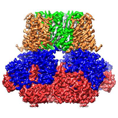 emd_8410.png emd_8410.png | 217.3 KB | ||
| Filedesc metadata | emd-8410.cif.gz | 6.8 KB | ||
| Archive directory |  http://ftp.pdbj.org/pub/emdb/structures/EMD-8410ftp://ftp.pdbj.org/pub/emdb/structures/EMD-8410 http://ftp.pdbj.org/pub/emdb/structures/EMD-8410ftp://ftp.pdbj.org/pub/emdb/structures/EMD-8410 | HTTPS FTP |
-Related structure data
| Related structure data |  5tj6MC M: atomic model generated by this map C: citing same article ( |
|---|---|
| Similar structure data |

-Links
| EMDB pages | EMDB (EBI/PDBe) / EMDataResource |
|---|---|
| Related items in Molecule of the Month |


-Map
| File | Download / File: emd_8410.map.gz / Format: CCP4 / Size: 64 MB / Type: IMAGE STORED AS FLOATING POINT NUMBER (4 BYTES) | ||||||||||||||||||||||||||||||||||||||||||||||||||||||||||||||||||||
|---|---|---|---|---|---|---|---|---|---|---|---|---|---|---|---|---|---|---|---|---|---|---|---|---|---|---|---|---|---|---|---|---|---|---|---|---|---|---|---|---|---|---|---|---|---|---|---|---|---|---|---|---|---|---|---|---|---|---|---|---|---|---|---|---|---|---|---|---|---|
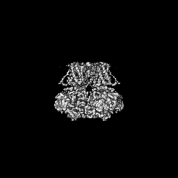
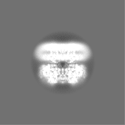
| Annotation | Mg and Ca-bound aplysia Slo1 | ||||||||||||||||||||||||||||||||||||||||||||||||||||||||||||||||||||
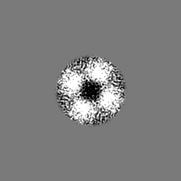
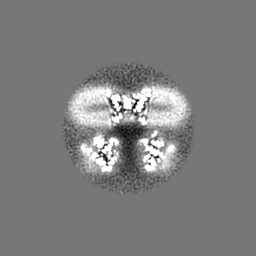
| Projections & slices | Image control
Images are generated by Spider. | ||||||||||||||||||||||||||||||||||||||||||||||||||||||||||||||||||||
| Voxel size | X=Y=Z: 1.3 Å | ||||||||||||||||||||||||||||||||||||||||||||||||||||||||||||||||||||
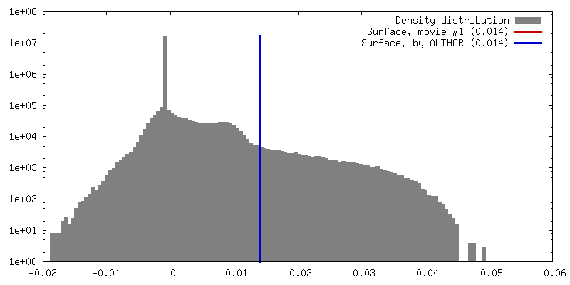
| Density |
| ||||||||||||||||||||||||||||||||||||||||||||||||||||||||||||||||||||
| Symmetry | Space group: 1 | ||||||||||||||||||||||||||||||||||||||||||||||||||||||||||||||||||||
| Details | EMDB XML:
CCP4 map header:
| ||||||||||||||||||||||||||||||||||||||||||||||||||||||||||||||||||||
 Z (Sec.)
 Y (Row.)
 X (Col.)


-Supplemental data
- Sample components
-Entire : Ca2+ bound aplysia Slo1
| Entire | Name: Ca2+ bound aplysia Slo1 |
|---|---|
| Components |
|
-Supramolecule #1: Ca2+ bound aplysia Slo1
| Supramolecule | Name: Ca2+ bound aplysia Slo1 / type: organelle_or_cellular_component / ID: 1 / Parent: 0 / Macromolecule list: #1 |
|---|---|
| Source (natural) | Organism: |
| Molecular weight | Theoretical: 400 KDa |
-Macromolecule #1: High conductance calcium-activated potassium channel
| Macromolecule | Name: High conductance calcium-activated potassium channel / type: protein_or_peptide / ID: 1 / Number of copies: 1 / Enantiomer: LEVO |
|---|---|
| Source (natural) | Organism: |
| Molecular weight | Theoretical: 120.308 KDa |
| Recombinant expression | Organism:  Trichoplusia ni (cabbage looper) Trichoplusia ni (cabbage looper) |
| Sequence | String: MASSSSTSCE PGDRQWYSFL ASSLVTFGSG LVVIIIYRIV LWLCCRKKKC IQVSNPVPTA RTTSLDQKSF MKNSDPEIGW MTEAKDWAG ELISGQTTTG RILVGLVFLL SIASLIIYFI DASTNTSVET CLPWSSSTTQ QVDLAFNVFF MIYFFIRFVA A NDKLWFWV ...String: MASSSSTSCE PGDRQWYSFL ASSLVTFGSG LVVIIIYRIV LWLCCRKKKC IQVSNPVPTA RTTSLDQKSF MKNSDPEIGW MTEAKDWAG ELISGQTTTG RILVGLVFLL SIASLIIYFI DASTNTSVET CLPWSSSTTQ QVDLAFNVFF MIYFFIRFVA A NDKLWFWV ELFSFVDYFT IPPSFVAIYL DRNWLGLRFL RALRLMSIPD ILTYLNVLKT STLIRLVQLV VSFVSLWLTA AG FLHLLEN SGDPFFDFGN AQHLTYWECL YFLMVTMSTV GFGDIFATTV LGRTFVVIFI MIFIGLFASF IPEIAEILGK RQK YGGSYK KERGKRHVVV CGYITFDSVS NFLKDFLHKD REDVDVEIVF LHKGLPGLEL EGLLKRHFTQ VEYFWGSVMD ANDL ERVKI QEADACLVLA NKYCQDPDQE DAANIMRVIS IKNYHSDIKV IVQLLQYHNK AYLLNIPSWD WKRGDDAVCV AELKL GFIA QSCLAPGFST LMANLFTMRS YKPTPEMSQW QTDYMRGTGM EMYTEYLSSA FNALTFPEAA ELCFSKLKLL LLAIEV RQE DTRESTLAIN PGPKVKIENA TQGFFIAESA EEVKRAFYYC KNCHANVSDV RQIKKCKCRP LAMFKKGAAA VLALQRT PG LAVEPDGEAN DKDKSRGTST SKAVTSFPEK RKPQSRRKPS TTLKSKSPSE DSVPPPPPPV DEPRKFDSTG MFHWCPDR P LNDCLQDRSQ ASASGLRNHV VVCLFADAAS PLIGLRNLVM PLRASNFHYH ELKPTIIVGN LDYLHREWKT LQNFPKLSI LPGSPLNRAN LRAVNINLCD MCVIVSAKDR NMEDPNLVDK EAILCSLNIK AMTFDDTMGL IQSSNFVPGG FSPLHENKRS QAGANVPLI TELANDSNVQ FLDQDDDDDP DTELYMTQPF ACGTAFAVSV LDSLMSTSYF NDNALTLIRT LITGGATPEL E QILAEGAG MRGGYCSPAV LANRDRCRVA QISLFDGPLA QFGQGGHYGE LFVYALRHFG ILCIGLYRFR DTNESVRSPS SK RYVITNP PEDFPLLPTD QVYVLTYKQI TNH UniProtKB: BK channel |
-Macromolecule #2: POTASSIUM ION
| Macromolecule | Name: POTASSIUM ION / type: ligand / ID: 2 / Number of copies: 5 / Formula: K |
|---|---|
| Molecular weight | Theoretical: 39.098 Da |
-Macromolecule #3: MAGNESIUM ION
| Macromolecule | Name: MAGNESIUM ION / type: ligand / ID: 3 / Number of copies: 1 / Formula: MG |
|---|---|
| Molecular weight | Theoretical: 24.305 Da |
-Macromolecule #4: CALCIUM ION
| Macromolecule | Name: CALCIUM ION / type: ligand / ID: 4 / Number of copies: 2 / Formula: CA |
|---|---|
| Molecular weight | Theoretical: 40.078 Da |
-Macromolecule #5: (1R)-2-{[(S)-{[(2S)-2,3-dihydroxypropyl]oxy}(hydroxy)phosphoryl]o...
| Macromolecule | Name: (1R)-2-{[(S)-{[(2S)-2,3-dihydroxypropyl]oxy}(hydroxy)phosphoryl]oxy}-1-[(hexadecanoyloxy)methyl]ethyl (9Z)-octadec-9-enoate type: ligand / ID: 5 / Number of copies: 15 / Formula: PGW |
|---|---|
| Molecular weight | Theoretical: 749.007 Da |
-Experimental details
-Structure determination
| Method | cryo EM |
|---|---|
 Processing Processing | single particle reconstruction |
| Aggregation state | particle |
-Sample preparation
| Concentration | 7 mg/mL | |||||||||||||||||||||||||||
|---|---|---|---|---|---|---|---|---|---|---|---|---|---|---|---|---|---|---|---|---|---|---|---|---|---|---|---|---|
| Buffer | pH: 8 Component:
| |||||||||||||||||||||||||||
| Grid | Model: Quantifoil R1.2/1.3 / Material: COPPER / Mesh: 400 / Support film - Material: CARBON / Support film - topology: HOLEY / Pretreatment - Type: GLOW DISCHARGE / Pretreatment - Time: 10 sec. / Pretreatment - Atmosphere: AIR | |||||||||||||||||||||||||||
| Vitrification | Cryogen name: ETHANE / Chamber humidity: 85 % / Chamber temperature: 293 K / Instrument: FEI VITROBOT MARK IV |
- Electron microscopy
| Microscope | FEI TITAN KRIOS |
|---|---|
| Image recording | Film or detector model: GATAN K2 SUMMIT (4k x 4k) / Detector mode: SUPER-RESOLUTION / Digitization - Dimensions - Width: 7420 pixel / Digitization - Dimensions - Height: 7676 pixel / Digitization - Frames/image: 1-50 / Number grids imaged: 1 / Number real images: 2000 / Average exposure time: 0.3 sec. / Average electron dose: 1.8 e/Å2 |
| Electron beam | Acceleration voltage: 300 kV / Electron source:  FIELD EMISSION GUN FIELD EMISSION GUN |
| Electron optics | Illumination mode: FLOOD BEAM / Imaging mode: BRIGHT FIELD / Cs: 2.7 mm / Nominal defocus max: 2.5 µm / Nominal defocus min: 1.0 µm / Nominal magnification: 22500 |
| Sample stage | Specimen holder model: FEI TITAN KRIOS AUTOGRID HOLDER / Cooling holder cryogen: NITROGEN |
| Experimental equipment |  Model: Titan Krios / Image courtesy: FEI Company |
+Image processing
-Atomic model buiding 1
| Refinement | Space: RECIPROCAL / Protocol: AB INITIO MODEL / Overall B value: 100 |
|---|---|
| Output model | PDB-5tj6: |
