 Movie
Movie Controller

[English] 日本語
 Yorodumi
- PDB-7sn4: Cryo-EM structure of the enterohemorrhagic E. coli O157:H7 flagel... -
+ Open data

- Basic information
| Entry | Database: PDB / ID: 7sn4 | |||||||||||||||||||||||||||
|---|---|---|---|---|---|---|---|---|---|---|---|---|---|---|---|---|---|---|---|---|---|---|---|---|---|---|---|---|
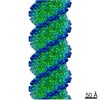
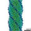
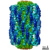
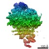
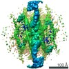
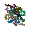
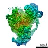
| Title | Cryo-EM structure of the enterohemorrhagic E. coli O157:H7 flagellar filament | |||||||||||||||||||||||||||
 Components Components | Flagellin | |||||||||||||||||||||||||||
 Keywords Keywords | STRUCTURAL PROTEIN / Bacteria flagellar filament / motility / flagellar polymorphism | |||||||||||||||||||||||||||
| Function / homology |  Function and homology information Function and homology informationbacterial-type flagellum / structural molecule activity / extracellular region Similarity search - Function | |||||||||||||||||||||||||||
| Biological species |  | |||||||||||||||||||||||||||
| Method | ELECTRON MICROSCOPY / helical reconstruction / cryo EM / Resolution: 3.6 Å | |||||||||||||||||||||||||||
 Authors Authors | Kreutzberger, M.A.B. / Wang, F. / Egelman, E.H. | |||||||||||||||||||||||||||
| Funding support | 1items
| |||||||||||||||||||||||||||
 Citation Citation | Journal: Nat Commun / Year: 2022 Title: Flagellin outer domain dimerization modulates motility in pathogenic and soil bacteria from viscous environments. Authors: Mark A B Kreutzberger / Richard C Sobe / Amber B Sauder / Sharanya Chatterjee / Alejandro Peña / Fengbin Wang / Jorge A Giron / Volker Kiessling / Tiago R D Costa / Vincent P Conticello / ...Authors: Mark A B Kreutzberger / Richard C Sobe / Amber B Sauder / Sharanya Chatterjee / Alejandro Peña / Fengbin Wang / Jorge A Giron / Volker Kiessling / Tiago R D Costa / Vincent P Conticello / Gad Frankel / Melissa M Kendall / Birgit E Scharf / Edward H Egelman /   Abstract: Flagellar filaments function as the propellers of the bacterial flagellum and their supercoiling is key to motility. The outer domains on the surface of the filament are non-critical for motility in ...Flagellar filaments function as the propellers of the bacterial flagellum and their supercoiling is key to motility. The outer domains on the surface of the filament are non-critical for motility in many bacteria and their structures and functions are not conserved. Here, we show the atomic cryo-electron microscopy structures for flagellar filaments from enterohemorrhagic Escherichia coli O157:H7, enteropathogenic E. coli O127:H6, Achromobacter, and Sinorhizobium meliloti, where the outer domains dimerize or tetramerize to form either a sheath or a screw-like surface. These dimers are formed by 180° rotations of half of the outer domains. The outer domain sheath (ODS) plays a role in bacterial motility by stabilizing an intermediate waveform and prolonging the tumbling of E. coli cells. Bacteria with these ODS and screw-like flagellar filaments are commonly found in soil and human intestinal environments of relatively high viscosity suggesting a role for the dimerization in these environments. | |||||||||||||||||||||||||||
| History |
|
- Structure visualization
| Movie |
Movie viewer |
|---|---|
| Structure viewer | Molecule: MolmilJmol/JSmol |
- Downloads & links
-Download
| PDBx/mmCIF format | 7sn4.cif.gz | 3.7 MB | Display | PDBx/mmCIF format |
|---|---|---|---|---|
| PDB format | pdb7sn4.ent.gz | Display | PDB format | |
| PDBx/mmJSON format | 7sn4.json.gz | Tree view | PDBx/mmJSON format | |
| Others |  Other downloads Other downloads |
-Validation report
| Arichive directory | https://data.pdbj.org/pub/pdb/validation_reports/sn/7sn4ftp://data.pdbj.org/pub/pdb/validation_reports/sn/7sn4 | HTTPS FTP |
|---|
-Related structure data
| Related structure data | 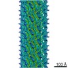 25211MC 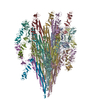 7sn7C 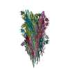 7sn9C 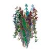 7sqdC 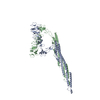 7sqjC M: map data used to model this data C: citing same article ( |
|---|---|
| Similar structure data |


-Links
 PDBj
PDBj- Assembly
| Deposited unit | 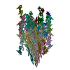
|
|---|---|
| 1 |
|
-Components
| #1: Protein | Mass: 60000.172 Da / Num. of mol.: 44 Source method: isolated from a genetically manipulated source Source: (gene. exp.) Has protein modification | N | |
|---|
-Experimental details
-Experiment
| Experiment | Method: ELECTRON MICROSCOPY |
|---|---|
| EM experiment | Aggregation state: FILAMENT / 3D reconstruction method: helical reconstruction |
- Sample preparation
| Component | Name: Bacterial flagellar filament / Type: COMPLEX / Entity ID: all / Source: NATURAL |
|---|---|
| Molecular weight | Experimental value: NO |
| Source (natural) | Organism: |
| Buffer solution | pH: 7.2 |
| Specimen | Embedding applied: NO / Shadowing applied: NO / Staining applied: NO / Vitrification applied: YES |
| Vitrification | Cryogen name: ETHANE |
- Electron microscopy imaging
| Experimental equipment |  Model: Titan Krios / Image courtesy: FEI Company |
|---|---|
| Microscopy | Model: TFS KRIOS |
| Electron gun | Electron source:  FIELD EMISSION GUN / Accelerating voltage: 300 kV / Illumination mode: FLOOD BEAM FIELD EMISSION GUN / Accelerating voltage: 300 kV / Illumination mode: FLOOD BEAM |
| Electron lens | Mode: BRIGHT FIELD |
| Image recording | Electron dose: 50 e/Å2 / Film or detector model: GATAN K3 (6k x 4k) |
- Processing
| Software |
| ||||||||||||||||||||||||
|---|---|---|---|---|---|---|---|---|---|---|---|---|---|---|---|---|---|---|---|---|---|---|---|---|---|
| CTF correction | Type: PHASE FLIPPING AND AMPLITUDE CORRECTION | ||||||||||||||||||||||||
| Helical symmerty | Angular rotation/subunit: 130.78 ° / Axial rise/subunit: 9.68 Å / Axial symmetry: C1 | ||||||||||||||||||||||||
| 3D reconstruction | Resolution: 3.6 Å / Resolution method: FSC 0.143 CUT-OFF / Num. of particles: 200000 / Symmetry type: HELICAL | ||||||||||||||||||||||||
| Refinement | Cross valid method: NONE Stereochemistry target values: GeoStd + Monomer Library + CDL v1.2 | ||||||||||||||||||||||||
| Displacement parameters | Biso mean: 150.11 Å2 | ||||||||||||||||||||||||
| Refine LS restraints |
|
