+ データを開く
- 基本情報
基本情報
| 登録情報 | データベース: PDB / ID: 7p1g | ||||||
|---|---|---|---|---|---|---|---|
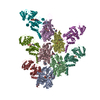
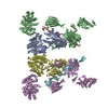
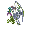
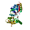
| タイトル | Structure of the P. aeruginosa ExoY-F-actin complex | ||||||
 要素 要素 |
| ||||||
 キーワード キーワード | TOXIN / Bacterial toxin / F-actin | ||||||
| 機能・相同性 |  機能・相同性情報 機能・相同性情報calcium- and calmodulin-responsive adenylate cyclase activity / cytoskeletal motor activator activity / tropomyosin binding / myosin heavy chain binding / detection of maltose stimulus / mesenchyme migration / troponin I binding / maltose transport complex / filamentous actin / actin filament bundle ...calcium- and calmodulin-responsive adenylate cyclase activity / cytoskeletal motor activator activity / tropomyosin binding / myosin heavy chain binding / detection of maltose stimulus / mesenchyme migration / troponin I binding / maltose transport complex / filamentous actin / actin filament bundle / maltose binding / carbohydrate transport / maltose transport / maltodextrin transmembrane transport / skeletal muscle thin filament assembly / actin filament bundle assembly / striated muscle thin filament / skeletal muscle myofibril / actin monomer binding / carbohydrate transmembrane transporter activity / ATP-binding cassette (ABC) transporter complex, substrate-binding subunit-containing / skeletal muscle fiber development / stress fiber / titin binding / actin filament polymerization / ATP-binding cassette (ABC) transporter complex / cell chemotaxis / filopodium / actin filament / 加水分解酵素; 酸無水物に作用; 酸無水物に作用・細胞または細胞小器官の運動に関与 / calcium-dependent protein binding / lamellipodium / outer membrane-bounded periplasmic space / cell body / periplasmic space / hydrolase activity / protein domain specific binding / DNA damage response / calcium ion binding / positive regulation of gene expression / magnesium ion binding / extracellular region / ATP binding / identical protein binding / membrane / cytoplasm 類似検索 - 分子機能 | ||||||
| 生物種 |   Pseudomonas aeruginosa PAO1 (緑膿菌) Pseudomonas aeruginosa PAO1 (緑膿菌)  Amanita phalloides (タマゴテングタケ) Amanita phalloides (タマゴテングタケ) | ||||||
| 手法 | 電子顕微鏡法 / 単粒子再構成法 / クライオ電子顕微鏡法 / 解像度: 3.2 Å | ||||||
 データ登録者 データ登録者 | Belyy, A. / Merino, F. / Raunser, S. | ||||||
| 資金援助 |  ドイツ, 1件 ドイツ, 1件
| ||||||
 引用 引用 |  ジャーナル: Nat Commun / 年: 2021 ジャーナル: Nat Commun / 年: 2021タイトル: Mechanism of actin-dependent activation of nucleotidyl cyclase toxins from bacterial human pathogens. 著者: Alexander Belyy / Felipe Merino / Undine Mechold / Stefan Raunser /   要旨: Bacterial human pathogens secrete initially inactive nucleotidyl cyclases that become potent enzymes by binding to actin inside eukaryotic host cells. The underlying molecular mechanism of this ...Bacterial human pathogens secrete initially inactive nucleotidyl cyclases that become potent enzymes by binding to actin inside eukaryotic host cells. The underlying molecular mechanism of this activation is, however, unclear. Here, we report structures of ExoY from Pseudomonas aeruginosa and Vibrio vulnificus bound to their corresponding activators F-actin and profilin-G-actin. The structures reveal that in contrast to the apo-state, two flexible regions become ordered and interact strongly with actin. The specific stabilization of these regions results in an allosteric stabilization of the nucleotide binding pocket and thereby to an activation of the enzyme. Differences in the sequence and conformation of the actin-binding regions are responsible for the selective binding to either F- or G-actin. Other nucleotidyl cyclase toxins that bind to calmodulin rather than actin undergo a similar disordered-to-ordered transition during activation, suggesting that the allosteric activation-by-stabilization mechanism of ExoY is conserved in these enzymes, albeit the different activator. | ||||||
| 履歴 |
|
- 構造の表示
構造の表示
| ムービー |
 ムービービューア ムービービューア |
|---|---|
| 構造ビューア | 分子:  Molmil Molmil Jmol/JSmol Jmol/JSmol |
- ダウンロードとリンク
- ダウンロード
| PDBx/mmCIF形式 |  7p1g.cif.gz 7p1g.cif.gz | 662.2 KB | 表示 |  PDBx/mmCIF形式 PDBx/mmCIF形式 |
|---|---|---|---|---|
| PDB形式 |  pdb7p1g.ent.gz pdb7p1g.ent.gz | 536.1 KB | 表示 |  PDB形式 PDB形式 |
| PDBx/mmJSON形式 |  7p1g.json.gz 7p1g.json.gz | ツリー表示 |  PDBx/mmJSON形式 PDBx/mmJSON形式 | |
| その他 |  その他のダウンロード その他のダウンロード |
-検証レポート
| 文書・要旨 |  7p1g_validation.pdf.gz 7p1g_validation.pdf.gz | 747.7 KB | 表示 |  wwPDB検証レポート wwPDB検証レポート |
|---|---|---|---|---|
| 文書・詳細版 |  7p1g_full_validation.pdf.gz 7p1g_full_validation.pdf.gz | 788.2 KB | 表示 | |
| XML形式データ |  7p1g_validation.xml.gz 7p1g_validation.xml.gz | 90.1 KB | 表示 | |
| CIF形式データ |  7p1g_validation.cif.gz 7p1g_validation.cif.gz | 132.8 KB | 表示 | |
| アーカイブディレクトリ |  https://data.pdbj.org/pub/pdb/validation_reports/p1/7p1g https://data.pdbj.org/pub/pdb/validation_reports/p1/7p1g ftp://data.pdbj.org/pub/pdb/validation_reports/p1/7p1g ftp://data.pdbj.org/pub/pdb/validation_reports/p1/7p1g | HTTPS FTP |
-関連構造データ
- リンク
リンク
- 集合体
集合体
| 登録構造単位 | 
|
|---|---|
| 1 |
|
- 要素
要素
-タンパク質 , 2種, 10分子 MKLNOCABDE
| #1: タンパク質 | 分子量: 84941.797 Da / 分子数: 5 / 由来タイプ: 組換発現 由来: (組換発現)   Pseudomonas aeruginosa PAO1 (緑膿菌) Pseudomonas aeruginosa PAO1 (緑膿菌)遺伝子: malE, b4034, JW3994, exoY, PA2191 / 発現宿主:  #2: タンパク質 | 分子量: 42096.953 Da / 分子数: 5 / 由来タイプ: 天然 / 由来: (天然)  |
|---|
-タンパク質・ペプチド , 1種, 5分子 HFGIJ
| #3: タンパク質・ペプチド | 分子量: 808.899 Da / 分子数: 5 / 由来タイプ: 合成 由来: (合成)  Amanita phalloides (タマゴテングタケ) Amanita phalloides (タマゴテングタケ) |
|---|
-非ポリマー , 4種, 25分子 


| #4: 化合物 | ChemComp-MG / #5: 化合物 | ChemComp-GH3 / #6: 化合物 | ChemComp-ADP / #7: 化合物 | ChemComp-PO4 / |
|---|
-詳細
| 研究の焦点であるリガンドがあるか | N |
|---|
-実験情報
-実験
| 実験 | 手法: 電子顕微鏡法 |
|---|---|
| EM実験 | 試料の集合状態: FILAMENT / 3次元再構成法: 単粒子再構成法 |
- 試料調製
試料調製
| 構成要素 | 名称: The complex of P. aeruginosa ExoY with F-actin / タイプ: COMPLEX / Entity ID: #1-#3 / 由来: MULTIPLE SOURCES |
|---|---|
| 分子量 | 実験値: NO |
| 緩衝液 | pH: 8 |
| 試料 | 包埋: NO / シャドウイング: NO / 染色: NO / 凍結: YES |
| 試料支持 | グリッドの材料: COPPER / グリッドのサイズ: 300 divisions/in. / グリッドのタイプ: Quantifoil R2/1 |
| 急速凍結 | 装置: FEI VITROBOT MARK III / 凍結剤: ETHANE / 湿度: 100 % |
- 電子顕微鏡撮影
| 実験機器 |  モデル: Titan Krios / 画像提供: FEI Company |
|---|---|
| 顕微鏡 | モデル: FEI TITAN KRIOS |
| 電子銃 | 電子線源:  FIELD EMISSION GUN / 加速電圧: 300 kV / 照射モード: SPOT SCAN FIELD EMISSION GUN / 加速電圧: 300 kV / 照射モード: SPOT SCAN |
| 電子レンズ | モード: BRIGHT FIELD / 最大 デフォーカス(公称値): 3500 nm / 最小 デフォーカス(公称値): 400 nm |
| 撮影 | 平均露光時間: 1.5 sec. / 電子線照射量: 93 e/Å2 / 検出モード: INTEGRATING フィルム・検出器のモデル: FEI FALCON III (4k x 4k) 撮影したグリッド数: 1 / 実像数: 12437 |
- 解析
解析
| EMソフトウェア |
| ||||||||||||||||||||||||||||||||||||
|---|---|---|---|---|---|---|---|---|---|---|---|---|---|---|---|---|---|---|---|---|---|---|---|---|---|---|---|---|---|---|---|---|---|---|---|---|---|
| CTF補正 | タイプ: PHASE FLIPPING AND AMPLITUDE CORRECTION | ||||||||||||||||||||||||||||||||||||
| 粒子像の選択 | 選択した粒子像数: 2249589 | ||||||||||||||||||||||||||||||||||||
| 対称性 | 点対称性: C1 (非対称) | ||||||||||||||||||||||||||||||||||||
| 3次元再構成 | 解像度: 3.2 Å / 解像度の算出法: FSC 0.143 CUT-OFF / 粒子像の数: 1535755 / アルゴリズム: FOURIER SPACE / 対称性のタイプ: POINT | ||||||||||||||||||||||||||||||||||||
| 原子モデル構築 | プロトコル: FLEXIBLE FIT / 空間: REAL | ||||||||||||||||||||||||||||||||||||
| 原子モデル構築 |
|
 ムービー
ムービー コントローラー


 PDBj
PDBj


