[English] 日本語
 Yorodumi
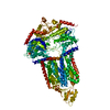
- PDB-7p04: Cryo-EM structure of Pdr5 from Saccharomyces cerevisiae in inward... -
+ Open data
- Basic information
| Entry | Database: PDB / ID: 7p04 | ||||||
|---|---|---|---|---|---|---|---|
| Title | Cryo-EM structure of Pdr5 from Saccharomyces cerevisiae in inward-facing conformation with ADP/ATP | ||||||
 Components Components | Pleiotropic ABC efflux transporter of multiple drugs | ||||||
 Keywords Keywords | TRANSPORT PROTEIN / ABC transporter / antibiotic resistance / membrane protein / fungal infection | ||||||
| Function / homology |  Function and homology information Function and homology informationintracellular monoatomic cation homeostasis / xenobiotic detoxification by transmembrane export across the plasma membrane / ABC-type xenobiotic transporter activity / response to cycloheximide / cell periphery / response to xenobiotic stimulus / response to antibiotic / ATP hydrolysis activity / mitochondrion / ATP binding ...intracellular monoatomic cation homeostasis / xenobiotic detoxification by transmembrane export across the plasma membrane / ABC-type xenobiotic transporter activity / response to cycloheximide / cell periphery / response to xenobiotic stimulus / response to antibiotic / ATP hydrolysis activity / mitochondrion / ATP binding / identical protein binding / plasma membrane Similarity search - Function | ||||||
| Biological species |  | ||||||
| Method | ELECTRON MICROSCOPY / single particle reconstruction / cryo EM / Resolution: 2.85 Å | ||||||
 Authors Authors | Szewczak-Harris, A. / Wagner, M. / Du, D. / Schmitt, L. / Luisi, B.F. | ||||||
| Funding support |  United Kingdom, 1items United Kingdom, 1items
| ||||||
 Citation Citation |  Journal: Nat Commun / Year: 2021 Journal: Nat Commun / Year: 2021Title: Structure and efflux mechanism of the yeast pleiotropic drug resistance transporter Pdr5. Authors: Andrzej Harris / Manuel Wagner / Dijun Du / Stefanie Raschka / Lea-Marie Nentwig / Holger Gohlke / Sander H J Smits / Ben F Luisi / Lutz Schmitt /    Abstract: Pdr5, a member of the extensive ABC transporter superfamily, is representative of a clinically relevant subgroup involved in pleiotropic drug resistance. Pdr5 and its homologues drive drug efflux ...Pdr5, a member of the extensive ABC transporter superfamily, is representative of a clinically relevant subgroup involved in pleiotropic drug resistance. Pdr5 and its homologues drive drug efflux through uncoupled hydrolysis of nucleotides, enabling organisms such as baker's yeast and pathogenic fungi to survive in the presence of chemically diverse antifungal agents. Here, we present the molecular structure of Pdr5 solved with single particle cryo-EM, revealing details of an ATP-driven conformational cycle, which mechanically drives drug translocation through an amphipathic channel, and a clamping switch within a conserved linker loop that acts as a nucleotide sensor. One half of the transporter remains nearly invariant throughout the cycle, while its partner undergoes changes that are transmitted across inter-domain interfaces to support a peristaltic motion of the pumped molecule. The efflux model proposed here rationalises the pleiotropic impact of Pdr5 and opens new avenues for the development of effective antifungal compounds. | ||||||
| History |
|
- Structure visualization
| Movie |
 Movie viewer Movie viewer |
|---|---|
| Structure viewer | Molecule:  Molmil Molmil Jmol/JSmol Jmol/JSmol |
- Downloads & links
- Download
| PDBx/mmCIF format |  7p04.cif.gz 7p04.cif.gz | 454.8 KB | Display |  PDBx/mmCIF format PDBx/mmCIF format |
|---|---|---|---|---|
| PDB format |  pdb7p04.ent.gz pdb7p04.ent.gz | 371.2 KB | Display |  PDB format PDB format |
| PDBx/mmJSON format |  7p04.json.gz 7p04.json.gz | Tree view |  PDBx/mmJSON format PDBx/mmJSON format | |
| Others |  Other downloads Other downloads |
-Validation report
| Arichive directory |  https://data.pdbj.org/pub/pdb/validation_reports/p0/7p04 https://data.pdbj.org/pub/pdb/validation_reports/p0/7p04 ftp://data.pdbj.org/pub/pdb/validation_reports/p0/7p04 ftp://data.pdbj.org/pub/pdb/validation_reports/p0/7p04 | HTTPS FTP |
|---|
-Related structure data
| Related structure data |  13143MC  7p03C  7p05C  7p06C M: map data used to model this data C: citing same article ( |
|---|---|
| Similar structure data |
- Links
Links
- Assembly
| Deposited unit | 
|
|---|---|
| 1 |
|
- Components
| #1: Protein | Mass: 170617.250 Da / Num. of mol.: 1 / Source method: isolated from a natural source Source: (natural)  Strain: ATCC 204508 / S288c / References: UniProt: P33302 |
|---|---|
| #2: Chemical | ChemComp-ATP / |
| #3: Chemical | ChemComp-ADP / |
| Has ligand of interest | Y |
| Has protein modification | Y |
-Experimental details
-Experiment
| Experiment | Method: ELECTRON MICROSCOPY |
|---|---|
| EM experiment | Aggregation state: PARTICLE / 3D reconstruction method: single particle reconstruction |
- Sample preparation
| Component | Name: Cryo-EM structure of Pdr5 from Saccharomyces cerevisiae in inward-facing conformation with ADP/ATP Type: COMPLEX / Entity ID: #1 / Source: NATURAL | |||||||||||||||
|---|---|---|---|---|---|---|---|---|---|---|---|---|---|---|---|---|
| Molecular weight | Experimental value: NO | |||||||||||||||
| Source (natural) | Organism:  | |||||||||||||||
| Buffer solution | pH: 7.8 | |||||||||||||||
| Buffer component |
| |||||||||||||||
| Specimen | Embedding applied: NO / Shadowing applied: NO / Staining applied: NO / Vitrification applied: YES | |||||||||||||||
| Specimen support | Grid material: COPPER / Grid mesh size: 300 divisions/in. / Grid type: Quantifoil R1.2/1.3 | |||||||||||||||
| Vitrification | Instrument: FEI VITROBOT MARK IV / Cryogen name: ETHANE / Humidity: 100 % / Chamber temperature: 277.15 K |
- Electron microscopy imaging
| Experimental equipment |  Model: Titan Krios / Image courtesy: FEI Company |
|---|---|
| Microscopy | Model: FEI TITAN KRIOS |
| Electron gun | Electron source:  FIELD EMISSION GUN / Accelerating voltage: 300 kV / Illumination mode: FLOOD BEAM FIELD EMISSION GUN / Accelerating voltage: 300 kV / Illumination mode: FLOOD BEAM |
| Electron lens | Mode: BRIGHT FIELD / Nominal magnification: 130000 X / Nominal defocus max: 2200 nm / Nominal defocus min: 700 nm / Cs: 2.7 mm / C2 aperture diameter: 50 µm |
| Specimen holder | Cryogen: HELIUM / Specimen holder model: FEI TITAN KRIOS AUTOGRID HOLDER |
| Image recording | Average exposure time: 1.31 sec. / Electron dose: 48.13 e/Å2 / Film or detector model: GATAN K3 (6k x 4k) |
| EM imaging optics | Energyfilter slit width: 20 eV |
- Processing
| Software | Name: PHENIX / Version: 1.18.2_3874: / Classification: refinement | ||||||||||||||||||||||||||||||||
|---|---|---|---|---|---|---|---|---|---|---|---|---|---|---|---|---|---|---|---|---|---|---|---|---|---|---|---|---|---|---|---|---|---|
| EM software |
| ||||||||||||||||||||||||||||||||
| CTF correction | Type: PHASE FLIPPING AND AMPLITUDE CORRECTION | ||||||||||||||||||||||||||||||||
| 3D reconstruction | Resolution: 2.85 Å / Resolution method: FSC 0.143 CUT-OFF / Num. of particles: 95283 / Algorithm: FOURIER SPACE / Num. of class averages: 1 / Symmetry type: POINT | ||||||||||||||||||||||||||||||||
| Atomic model building | Protocol: AB INITIO MODEL | ||||||||||||||||||||||||||||||||
| Refine LS restraints |
|
 Movie
Movie Controller


 PDBj
PDBj


