 Movie
Movie Controller

[English] 日本語
 Yorodumi
- PDB-7ovt: major seeded in vitro fibril morphology from murine SAA1.1 protein -
+ Open data

- Basic information
| Entry | Database: PDB / ID: 7ovt | |||||||||||||||||||||
|---|---|---|---|---|---|---|---|---|---|---|---|---|---|---|---|---|---|---|---|---|---|---|
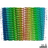
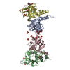
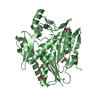
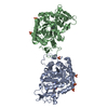
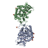
| Title | major seeded in vitro fibril morphology from murine SAA1.1 protein | |||||||||||||||||||||
 Components Components | Serum amyloid A-2 protein | |||||||||||||||||||||
 Keywords Keywords | PROTEIN FIBRIL / systemic amyloidosis / seeded / misfolding disease / inflammation / prion | |||||||||||||||||||||
| Function / homology |  Function and homology information Function and homology informationresponse to stilbenoid / high-density lipoprotein particle / cytoplasmic microtubule / acute-phase response / G protein-coupled receptor binding Similarity search - Function | |||||||||||||||||||||
| Biological species |  | |||||||||||||||||||||
| Method | ELECTRON MICROSCOPY / helical reconstruction / cryo EM / Resolution: 2.69 Å | |||||||||||||||||||||
 Authors Authors | Heerde, T. / Schmidt, M. / Faendrich, M. | |||||||||||||||||||||
| Funding support |  Germany, 6items Germany, 6items
| |||||||||||||||||||||
 Citation Citation | Journal: Nat Commun / Year: 2022 Title: Cryo-EM demonstrates the in vitro proliferation of an ex vivo amyloid fibril morphology by seeding. Authors: Thomas Heerde / Matthies Rennegarbe / Alexander Biedermann / Dilan Savran / Peter B Pfeiffer / Manuel Hitzenberger / Julian Baur / Ioana Puscalau-Girtu / Martin Zacharias / Nadine Schwierz / ...Authors: Thomas Heerde / Matthies Rennegarbe / Alexander Biedermann / Dilan Savran / Peter B Pfeiffer / Manuel Hitzenberger / Julian Baur / Ioana Puscalau-Girtu / Martin Zacharias / Nadine Schwierz / Christian Haupt / Matthias Schmidt / Marcus Fändrich / Abstract: Several studies showed that seeding of solutions of monomeric fibril proteins with ex vivo amyloid fibrils accelerated the kinetics of fibril formation in vitro but did not necessarily replicate the ...Several studies showed that seeding of solutions of monomeric fibril proteins with ex vivo amyloid fibrils accelerated the kinetics of fibril formation in vitro but did not necessarily replicate the seed structure. In this research we use cryo-electron microscopy and other methods to analyze the ability of serum amyloid A (SAA)1.1-derived amyloid fibrils, purified from systemic AA amyloidosis tissue, to seed solutions of recombinant SAA1.1 protein. We show that 98% of the seeded fibrils remodel the full fibril structure of the main ex vivo fibril morphology, which we used for seeding, while they are notably different from unseeded in vitro fibrils. The seeded fibrils show a similar proteinase K resistance as ex vivo fibrils and are substantially more stable to proteolytic digestion than unseeded in vitro fibrils. Our data support the view that the fibril morphology contributes to determining proteolytic stability and that pathogenic amyloid fibrils arise from proteolytic selection. | |||||||||||||||||||||
| History |
|
- Structure visualization
| Movie |
Movie viewer |
|---|---|
| Structure viewer | Molecule: MolmilJmol/JSmol |
- Downloads & links
-Download
| PDBx/mmCIF format | 7ovt.cif.gz | 150 KB | Display | PDBx/mmCIF format |
|---|---|---|---|---|
| PDB format | pdb7ovt.ent.gz | 120.8 KB | Display | PDB format |
| PDBx/mmJSON format | 7ovt.json.gz | Tree view | PDBx/mmJSON format | |
| Others |  Other downloads Other downloads |
-Validation report
| Arichive directory | https://data.pdbj.org/pub/pdb/validation_reports/ov/7ovtftp://data.pdbj.org/pub/pdb/validation_reports/ov/7ovt | HTTPS FTP |
|---|
-Related structure data
| Related structure data | 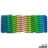 13089MC M: map data used to model this data C: citing same article ( |
|---|---|
| Similar structure data |


-Links
 PDBj
PDBj

- Assembly
| Deposited unit | 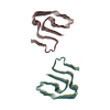
|
|---|---|
| 1 |
|
-Components
| #1: Protein | Mass: 11622.629 Da / Num. of mol.: 12 Source method: isolated from a genetically manipulated source Details: amyloid fibril / Source: (gene. exp.)  |
|---|
-Experimental details
-Experiment
| Experiment | Method: ELECTRON MICROSCOPY |
|---|---|
| EM experiment | Aggregation state: HELICAL ARRAY / 3D reconstruction method: helical reconstruction |
- Sample preparation
| Component | Name: Murine serum amyloid A1 (SAA1) amyloid fibril / Type: COMPLEX Details: in vitro murine SAA amyloid fibril morphology i; Seeded with ex vivo material Entity ID: all / Source: RECOMBINANT |
|---|---|
| Molecular weight | Units: KILODALTONS/NANOMETER / Experimental value: NO |
| Source (natural) | Organism: |
| Source (recombinant) | Organism: |
| Buffer solution | pH: 8.5 / Details: 10mM Tris(hydroxymethyl)aminomethane (Tris) |
| Buffer component | Conc.: 10 mM / Name: Tris(hydroxymethyl)aminomethane / Formula: (HOCH2)3CNH2 |
| Specimen | Conc.: 0.2 mg/ml / Embedding applied: NO / Shadowing applied: NO / Staining applied: NO / Vitrification applied: YES |
| Specimen support | Grid material: COPPER / Grid mesh size: 400 divisions/in. / Grid type: C-flat-1.2/1.3 |
| Vitrification | Instrument: FEI VITROBOT MARK III / Cryogen name: ETHANE / Humidity: 96 % |
- Electron microscopy imaging
| Experimental equipment |  Model: Titan Krios / Image courtesy: FEI Company |
|---|---|
| Microscopy | Model: FEI TITAN KRIOS |
| Electron gun | Electron source:  FIELD EMISSION GUN / Accelerating voltage: 300 kV / Illumination mode: FLOOD BEAM FIELD EMISSION GUN / Accelerating voltage: 300 kV / Illumination mode: FLOOD BEAM |
| Electron lens | Mode: BRIGHT FIELD / Cs: 2.7 mm |
| Specimen holder | Cryogen: NITROGEN |
| Image recording | Average exposure time: 12 sec. / Electron dose: 40 e/Å2 / Detector mode: COUNTING / Film or detector model: GATAN K2 SUMMIT (4k x 4k) |
| Image scans | Movie frames/image: 40 |
- Processing
| EM software |
| ||||||||||||||||||||||||||||
|---|---|---|---|---|---|---|---|---|---|---|---|---|---|---|---|---|---|---|---|---|---|---|---|---|---|---|---|---|---|
| CTF correction | Type: PHASE FLIPPING AND AMPLITUDE CORRECTION | ||||||||||||||||||||||||||||
| Helical symmerty | Angular rotation/subunit: 179.425 ° / Axial rise/subunit: 2.4 Å / Axial symmetry: C1 | ||||||||||||||||||||||||||||
| Particle selection | Num. of particles selected: 141159 | ||||||||||||||||||||||||||||
| 3D reconstruction | Resolution: 2.69 Å / Resolution method: FSC 0.143 CUT-OFF / Num. of particles: 107856 / Symmetry type: HELICAL | ||||||||||||||||||||||||||||
| Atomic model building | Protocol: BACKBONE TRACE / Space: REAL / Target criteria: correlation coefficient |
