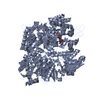
Movie
Movie Controller

[English] 日本語
 Yorodumi
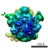
- PDB-7mft: Glutamate synthase, glutamate dehydrogenase counter-enzyme comple... -
+ Open data

- Basic information
| Entry | Database: PDB / ID: 7mft | |||||||||
|---|---|---|---|---|---|---|---|---|---|---|
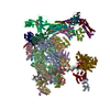
| Title | Glutamate synthase, glutamate dehydrogenase counter-enzyme complex (GudB6-GltA6-GltB6) | |||||||||
 Components Components |
| |||||||||
 Keywords Keywords | CYTOSOLIC PROTEIN / Counter-enzyme complex / glutamate synthase / glutamate dehydrogenae | |||||||||
| Function / homology |  Function and homology information Function and homology informationglutamate synthase activity / oxidoreductase activity, acting on the CH-NH2 group of donors, NAD or NADP as acceptor / L-glutamate dehydrogenase (NAD+) activity / L-glutamate catabolic process / : / L-glutamine metabolic process / 3 iron, 4 sulfur cluster binding / iron-sulfur cluster binding / nucleotide binding / metal ion binding Similarity search - Function | |||||||||
| Biological species |  | |||||||||
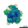
| Method | ELECTRON MICROSCOPY / single particle reconstruction / cryo EM / Resolution: 3.9 Å | |||||||||
 Authors Authors | Jayaraman, V. / Lee, D.J. / Elad, N. / Fraser, J.S. / Tawfik, D.S. | |||||||||
| Funding support |  Israel, Israel,  United States, 2items United States, 2items
| |||||||||
 Citation Citation | Journal: Nat Chem Biol / Year: 2022 Title: A counter-enzyme complex regulates glutamate metabolism in Bacillus subtilis. Authors: Vijay Jayaraman / D John Lee / Nadav Elad / Shay Vimer / Michal Sharon / James S Fraser / Dan S Tawfik / Abstract: Multi-enzyme assemblies composed of metabolic enzymes catalyzing sequential reactions are being increasingly studied. Here, we report the discovery of a 1.6 megadalton multi-enzyme complex from ...Multi-enzyme assemblies composed of metabolic enzymes catalyzing sequential reactions are being increasingly studied. Here, we report the discovery of a 1.6 megadalton multi-enzyme complex from Bacillus subtilis composed of two enzymes catalyzing opposite ('counter-enzymes') rather than sequential reactions: glutamate synthase (GltAB) and glutamate dehydrogenase (GudB), which make and break glutamate, respectively. In vivo and in vitro studies show that the primary role of complex formation is to inhibit the activity of GudB. Using cryo-electron microscopy, we elucidated the structure of the complex and the molecular basis of inhibition of GudB by GltAB. The complex exhibits unusual oscillatory progress curves and is necessary for both planktonic growth, in glutamate-limiting conditions, and for biofilm growth, in glutamate-rich media. The regulation of a key metabolic enzyme by complexing with its counter enzyme may thus enable cell growth under fluctuating glutamate concentrations. | |||||||||
| History |
|
- Structure visualization
| Movie |
Movie viewer |
|---|---|
| Structure viewer | Molecule: MolmilJmol/JSmol |
- Downloads & links
-Download
| PDBx/mmCIF format | 7mft.cif.gz | 786.3 KB | Display | PDBx/mmCIF format |
|---|---|---|---|---|
| PDB format | pdb7mft.ent.gz | 646.4 KB | Display | PDB format |
| PDBx/mmJSON format | 7mft.json.gz | Tree view | PDBx/mmJSON format | |
| Others |  Other downloads Other downloads |
-Validation report
| Arichive directory | https://data.pdbj.org/pub/pdb/validation_reports/mf/7mftftp://data.pdbj.org/pub/pdb/validation_reports/mf/7mft | HTTPS FTP |
|---|
-Related structure data
| Related structure data |  23825MC  7mfmC C: citing same article ( M: map data used to model this data |
|---|---|
| Similar structure data |


-Links
 PDBj
PDBj


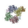
- Assembly
| Deposited unit | 
|
|---|---|
| 1 | x 6
|
| 2 |
|
| 3 | 
|
| Symmetry | Point symmetry: (Schoenflies symbol: D3 (2x3 fold dihedral)) |
-Components
-Protein , 1 types, 1 molecules A
| #1: Protein | Mass: 47100.715 Da / Num. of mol.: 1 Source method: isolated from a genetically manipulated source Source: (gene. exp.) Gene: gudB, B4122_2172, ETA10_11605, ETK61_12750, GII81_12565, SC09_Contig24orf00413 Production host: |
|---|
-Glutamate synthase (NADPH) ... , 2 types, 2 molecules GI
| #2: Protein | Mass: 169006.375 Da / Num. of mol.: 1 Source method: isolated from a genetically manipulated source Source: (gene. exp.) Gene: B4417_0011 / Production host: |
|---|---|
| #3: Protein | Mass: 58010.598 Da / Num. of mol.: 1 / Mutation: D78G Source method: isolated from a genetically manipulated source Source: (gene. exp.) Gene: B4417_0010 / Production host: |
-Non-polymers , 4 types, 5 molecules 


| #4: Chemical | ChemComp-FMN /  Mass: 456.344 Da / Num. of mol.: 1 / Source method: obtained synthetically / Formula: C17H21N4O9P Mass: 456.344 Da / Num. of mol.: 1 / Source method: obtained synthetically / Formula: C17H21N4O9P |
|---|---|
| #5: Chemical | ChemComp-F3S /  Mass: 295.795 Da / Num. of mol.: 1 / Source method: obtained synthetically / Formula: Fe3S4 Mass: 295.795 Da / Num. of mol.: 1 / Source method: obtained synthetically / Formula: Fe3S4 |
| #6: Chemical | ChemComp-FAD /  Mass: 785.550 Da / Num. of mol.: 1 / Source method: obtained synthetically / Formula: C27H33N9O15P2 / Comment: FAD*YM Mass: 785.550 Da / Num. of mol.: 1 / Source method: obtained synthetically / Formula: C27H33N9O15P2 / Comment: FAD*YM |
| #7: Chemical |  Mass: 351.640 Da / Num. of mol.: 2 / Source method: obtained synthetically / Formula: Fe4S4 Mass: 351.640 Da / Num. of mol.: 2 / Source method: obtained synthetically / Formula: Fe4S4 |
-Details
| Has ligand of interest | N |
|---|
-Experimental details
-Experiment
| Experiment | Method: ELECTRON MICROSCOPY |
|---|---|
| EM experiment | Aggregation state: PARTICLE / 3D reconstruction method: single particle reconstruction |
- Sample preparation
| Component | Name: GudB-GltA-GltB / Type: COMPLEX Details: Asymmetric unit (GudB-GltA-GltB) from a full GudB6-GltA6-GltB6 complex. Entity ID: #1-#3 / Source: RECOMBINANT |
|---|---|
| Molecular weight | Value: 1.62 MDa / Experimental value: YES |
| Source (natural) | Organism: |
| Source (recombinant) | Organism: |
| Buffer solution | pH: 7.9 |
| Specimen | Conc.: 0.5 mg/ml / Embedding applied: NO / Shadowing applied: NO / Staining applied: NO / Vitrification applied: YES |
| Vitrification | Cryogen name: ETHANE |
- Electron microscopy imaging
| Experimental equipment |  Model: Titan Krios / Image courtesy: FEI Company |
|---|---|
| Microscopy | Model: FEI TITAN KRIOS |
| Electron gun | Electron source:  FIELD EMISSION GUN / Accelerating voltage: 300 kV / Illumination mode: FLOOD BEAM FIELD EMISSION GUN / Accelerating voltage: 300 kV / Illumination mode: FLOOD BEAM |
| Electron lens | Mode: BRIGHT FIELD |
| Specimen holder | Cryogen: NITROGEN / Specimen holder model: FEI TITAN KRIOS AUTOGRID HOLDER |
| Image recording | Electron dose: 47.7 e/Å2 / Film or detector model: GATAN K3 BIOQUANTUM (6k x 4k) |
- Processing
| Software | Name: PHENIX / Version: 1.19.1_4122: / Classification: refinement | |||||||||||||||||||||||||
|---|---|---|---|---|---|---|---|---|---|---|---|---|---|---|---|---|---|---|---|---|---|---|---|---|---|---|
| EM software |
| |||||||||||||||||||||||||
| CTF correction | Type: PHASE FLIPPING AND AMPLITUDE CORRECTION | |||||||||||||||||||||||||
| Particle selection | Num. of particles selected: 60487 | |||||||||||||||||||||||||
| 3D reconstruction | Resolution: 3.9 Å / Resolution method: FSC 0.143 CUT-OFF / Num. of particles: 11878 / Symmetry type: POINT | |||||||||||||||||||||||||
| Atomic model building | Protocol: RIGID BODY FIT / Space: REAL Details: Real-space refinement involved iterative rounds of ISOLDE, Coot and PHENIX refinement. 1OFD and 6S6T were used as templates for homology modeling in SWISS-MODEL. | |||||||||||||||||||||||||
| Atomic model building | 3D fitting-ID: 1 / Source name: PDB / Type: experimental model
| |||||||||||||||||||||||||
| Refinement | Cross valid method: NONE Stereochemistry target values: GeoStd + Monomer Library + CDL v1.2 | |||||||||||||||||||||||||
| Displacement parameters | Biso mean: 97.66 Å2 | |||||||||||||||||||||||||
| Refine LS restraints |
|