 Movie
Movie Controller

[English] 日本語
 Yorodumi
- PDB-7jw1: Satellite phage P4 procapsid including size determination (Sid) p... -
+ Open data

- Basic information
| Entry | Database: PDB / ID: 7jw1 | |||||||||||||||||||||||||||||||||||||||||||||||||||||||||||||||
|---|---|---|---|---|---|---|---|---|---|---|---|---|---|---|---|---|---|---|---|---|---|---|---|---|---|---|---|---|---|---|---|---|---|---|---|---|---|---|---|---|---|---|---|---|---|---|---|---|---|---|---|---|---|---|---|---|---|---|---|---|---|---|---|---|
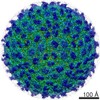
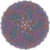
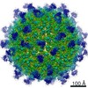
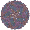
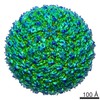
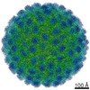
| Title | Satellite phage P4 procapsid including size determination (Sid) protein | |||||||||||||||||||||||||||||||||||||||||||||||||||||||||||||||
 Components Components |
| |||||||||||||||||||||||||||||||||||||||||||||||||||||||||||||||
 Keywords Keywords | VIRUS / bacteriophage / phage / procapsid / satellite phage / size determination / capsid protein / molecular piracy | |||||||||||||||||||||||||||||||||||||||||||||||||||||||||||||||
| Function / homology | Bacteriophage P2, capsid / Phage major capsid protein, P2 family / viral capsid / Glycoprotein 3 / Capsid proteins Function and homology information Function and homology information | |||||||||||||||||||||||||||||||||||||||||||||||||||||||||||||||
| Biological species |  Escherichia phage P2 (virus)Enterobacteria phage P4 (virus) Escherichia phage P2 (virus)Enterobacteria phage P4 (virus) | |||||||||||||||||||||||||||||||||||||||||||||||||||||||||||||||
| Method | ELECTRON MICROSCOPY / single particle reconstruction / cryo EM / Resolution: 4.19 Å | |||||||||||||||||||||||||||||||||||||||||||||||||||||||||||||||
 Authors Authors | Kizziah, J.L. / Dokland, T. | |||||||||||||||||||||||||||||||||||||||||||||||||||||||||||||||
| Funding support |  United States, 1items United States, 1items
| |||||||||||||||||||||||||||||||||||||||||||||||||||||||||||||||
 Citation Citation | Journal: Viruses / Year: 2020 Title: Structure of the Capsid Size-Determining Scaffold of "Satellite" Bacteriophage P4. Authors: James L Kizziah / Cynthia M Rodenburg / Terje Dokland / Abstract: P4 is a mobile genetic element (MGE) that can exist as a plasmid or integrated into its host genome, but becomes packaged into phage particles by a helper bacteriophage, such as P2. P4 is the ...P4 is a mobile genetic element (MGE) that can exist as a plasmid or integrated into its host genome, but becomes packaged into phage particles by a helper bacteriophage, such as P2. P4 is the original example of what we have termed "molecular piracy", the process by which one MGE usurps the life cycle of another for its own propagation. The P2 helper provides most of the structural gene products for assembly of the P4 virion. However, when P4 is mobilized by P2, the resulting capsids are smaller than those normally formed by P2 alone. The P4-encoded protein responsible for this size change is called Sid, which forms an external scaffolding cage around the P4 procapsids. We have determined the high-resolution structure of P4 procapsids, allowing us to build an atomic model for Sid as well as the gpN capsid protein. Sixty copies of Sid form an intertwined dodecahedral cage around the = 4 procapsid, making contact with only one out of the four symmetrically non-equivalent copies of gpN. Our structure provides a basis for understanding the mutants in gpN that prevent small capsid formation, as well as the "super-sid" mutations that counteract the effect of the mutations, and suggests a model for capsid size redirection by Sid. | |||||||||||||||||||||||||||||||||||||||||||||||||||||||||||||||
| History |
|
- Structure visualization
| Movie |
Movie viewer |
|---|---|
| Structure viewer | Molecule: MolmilJmol/JSmol |
- Downloads & links
-Download
| PDBx/mmCIF format | 7jw1.cif.gz | 497.9 KB | Display | PDBx/mmCIF format |
|---|---|---|---|---|
| PDB format | pdb7jw1.ent.gz | 413.5 KB | Display | PDB format |
| PDBx/mmJSON format | 7jw1.json.gz | Tree view | PDBx/mmJSON format | |
| Others |  Other downloads Other downloads |
-Validation report
| Arichive directory | https://data.pdbj.org/pub/pdb/validation_reports/jw/7jw1ftp://data.pdbj.org/pub/pdb/validation_reports/jw/7jw1 | HTTPS FTP |
|---|
-Related structure data
| Related structure data |  22513MC M: map data used to model this data C: citing same article ( |
|---|---|
| Similar structure data |


-Links
 PDBj
PDBj- Assembly
| Deposited unit | 
|
|---|---|
| 1 | x 30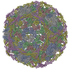
|
| 2 |
|
| Symmetry | Point symmetry: (Schoenflies symbol: I (icosahedral)) |
-Components
| #1: Protein | Mass: 40291.484 Da / Num. of mol.: 8 Source method: isolated from a genetically manipulated source Source: (gene. exp.) Escherichia phage P2 (virus) / Production host:  #2: Protein | Mass: 27296.814 Da / Num. of mol.: 2 Source method: isolated from a genetically manipulated source Source: (gene. exp.) Enterobacteria phage P4 (virus) / Gene: sid / Production host: Has protein modification | Y | |
|---|
-Experimental details
-Experiment
| Experiment | Method: ELECTRON MICROSCOPY |
|---|---|
| EM experiment | Aggregation state: PARTICLE / 3D reconstruction method: single particle reconstruction |
- Sample preparation
| Component | Name: P4 procapsid / Type: COMPLEX / Entity ID: all / Source: RECOMBINANT |
|---|---|
| Molecular weight | Experimental value: NO |
| Source (natural) | Organism: |
| Source (recombinant) | Organism: |
| Buffer solution | pH: 7.4 |
| Specimen | Embedding applied: NO / Shadowing applied: NO / Staining applied: NO / Vitrification applied: YES |
| Vitrification | Cryogen name: ETHANE |
- Electron microscopy imaging
| Experimental equipment |  Model: Titan Krios / Image courtesy: FEI Company |
|---|---|
| Microscopy | Model: FEI TITAN KRIOS |
| Electron gun | Electron source:  FIELD EMISSION GUN / Accelerating voltage: 300 kV / Illumination mode: FLOOD BEAM FIELD EMISSION GUN / Accelerating voltage: 300 kV / Illumination mode: FLOOD BEAM |
| Electron lens | Mode: BRIGHT FIELD |
| Image recording | Electron dose: 30 e/Å2 / Film or detector model: GATAN K3 (6k x 4k) |
- Processing
| Software | Name: PHENIX / Version: 1.17.1_3660: / Classification: refinement | ||||||||||||||||||||||||
|---|---|---|---|---|---|---|---|---|---|---|---|---|---|---|---|---|---|---|---|---|---|---|---|---|---|
| EM software | Name: PHENIX / Category: model refinement | ||||||||||||||||||||||||
| CTF correction | Type: PHASE FLIPPING ONLY | ||||||||||||||||||||||||
| 3D reconstruction | Resolution: 4.19 Å / Resolution method: FSC 0.143 CUT-OFF / Num. of particles: 438018 / Symmetry type: POINT | ||||||||||||||||||||||||
| Refine LS restraints |
|
