 Movie
Movie Controller


+ Open data

- Basic information
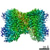
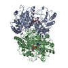
| Entry | Database: PDB / ID: 7jsk | ||||||||||||||||||||||||
|---|---|---|---|---|---|---|---|---|---|---|---|---|---|---|---|---|---|---|---|---|---|---|---|---|---|
| Title | Structure of the NaCT-Citrate complex | ||||||||||||||||||||||||
 Components Components | Solute carrier family 13 member 5 | ||||||||||||||||||||||||
 Keywords Keywords | MEMBRANE PROTEIN / Transporter | ||||||||||||||||||||||||
| Function / homology |  Function and homology information Function and homology informationorganic acid:sodium symporter activity / fumarate transport / oxaloacetate transport / succinate transport / sodium:dicarboxylate symporter activity / citrate transmembrane transporter activity / citrate transport / Sodium-coupled sulphate, di- and tri-carboxylate transporters / alpha-ketoglutarate transport / succinate transmembrane transporter activity ...organic acid:sodium symporter activity / fumarate transport / oxaloacetate transport / succinate transport / sodium:dicarboxylate symporter activity / citrate transmembrane transporter activity / citrate transport / Sodium-coupled sulphate, di- and tri-carboxylate transporters / alpha-ketoglutarate transport / succinate transmembrane transporter activity / cellular response to lithium ion / transmembrane transport / nucleoplasm / identical protein binding / plasma membrane / cytosol Similarity search - Function | ||||||||||||||||||||||||
| Biological species |  Homo sapiens (human) Homo sapiens (human) | ||||||||||||||||||||||||
| Method | ELECTRON MICROSCOPY / single particle reconstruction / cryo EM / Resolution: 3.04 Å | ||||||||||||||||||||||||
 Authors Authors | Sauer, D.B. / Wang, B. / Song, J. / Rice, W.J. / Wang, D.N. | ||||||||||||||||||||||||
| Funding support |  United States, 7items United States, 7items
| ||||||||||||||||||||||||
 Citation Citation | Journal: Nature / Year: 2021 Title: Structure and inhibition mechanism of the human citrate transporter NaCT. Authors: David B Sauer / Jinmei Song / Bing Wang / Jacob K Hilton / Nathan K Karpowich / Joseph A Mindell / William J Rice / Da-Neng Wang / Abstract: Citrate is best known as an intermediate in the tricarboxylic acid cycle of the cell. In addition to this essential role in energy metabolism, the tricarboxylate anion also acts as both a precursor ...Citrate is best known as an intermediate in the tricarboxylic acid cycle of the cell. In addition to this essential role in energy metabolism, the tricarboxylate anion also acts as both a precursor and a regulator of fatty acid synthesis. Thus, the rate of fatty acid synthesis correlates directly with the cytosolic concentration of citrate. Liver cells import citrate through the sodium-dependent citrate transporter NaCT (encoded by SLC13A5) and, as a consequence, this protein is a potential target for anti-obesity drugs. Here, to understand the structural basis of its inhibition mechanism, we determined cryo-electron microscopy structures of human NaCT in complexes with citrate or a small-molecule inhibitor. These structures reveal how the inhibitor-which binds to the same site as citrate-arrests the transport cycle of NaCT. The NaCT-inhibitor structure also explains why the compound selectively inhibits NaCT over two homologous human dicarboxylate transporters, and suggests ways to further improve the affinity and selectivity. Finally, the NaCT structures provide a framework for understanding how various mutations abolish the transport activity of NaCT in the brain and thereby cause epilepsy associated with mutations in SLC13A5 in newborns (which is known as SLC13A5-epilepsy). | ||||||||||||||||||||||||
| History |
|
- Structure visualization
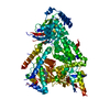
| Movie |
Movie viewer |
|---|---|
| Structure viewer | Molecule: MolmilJmol/JSmol |
- Downloads & links
-Download
| PDBx/mmCIF format | 7jsk.cif.gz | 319.1 KB | Display | PDBx/mmCIF format |
|---|---|---|---|---|
| PDB format | pdb7jsk.ent.gz | 260.6 KB | Display | PDB format |
| PDBx/mmJSON format | 7jsk.json.gz | Tree view | PDBx/mmJSON format | |
| Others |  Other downloads Other downloads |
-Validation report
| Arichive directory | https://data.pdbj.org/pub/pdb/validation_reports/js/7jskftp://data.pdbj.org/pub/pdb/validation_reports/js/7jsk | HTTPS FTP |
|---|
-Related structure data
| Related structure data |  22457MC 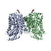 7jsjC M: map data used to model this data C: citing same article ( |
|---|---|
| Similar structure data | |
| EM raw data | EMPIAR-10728 (Title: Micrographs of the NaCT-Citrate complex / Data size: 2.4 TB Data #1: Unaligned 0 degree tilt micrographs of NaCT-Citrate data collection 29jun2020 [micrographs - multiframe] Data #2: Unaligned 20 degree tilt micrographs of NaCT-Citrate data collection 29jun2020 [micrographs - multiframe] Data #3: Unaligned 40 degree tilt micrographs of NaCT-Citrate data collection 29jun2020 [micrographs - multiframe] Data #4: Unaligned 50 degree tilt micrographs of NaCT-Citrate data collection 04jul2020 [micrographs - multiframe] Data #5: Unaligned 40 degree tilt micrographs of NaCT-Citrate data collection 04jul2020 [micrographs - multiframe]) |


-Links
 PDBj
PDBj- Assembly
| Deposited unit | 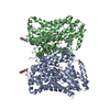
|
|---|---|
| 1 |
|
-Components
| #1: Protein | Mass: 63110.812 Da / Num. of mol.: 2 Source method: isolated from a genetically manipulated source Source: (gene. exp.) Homo sapiens (human) / Gene: SLC13A5, NACT / Production host:  Trichoplusia ni (cabbage looper) / References: UniProt: Q86YT5 Trichoplusia ni (cabbage looper) / References: UniProt: Q86YT5#2: Sugar |   Type: D-saccharide, beta linking / Mass: 221.208 Da / Num. of mol.: 2 / Source method: obtained synthetically / Formula: C8H15NO6 Type: D-saccharide, beta linking / Mass: 221.208 Da / Num. of mol.: 2 / Source method: obtained synthetically / Formula: C8H15NO6#3: Chemical |   Mass: 192.124 Da / Num. of mol.: 2 / Source method: obtained synthetically / Formula: C6H8O7 / Feature type: SUBJECT OF INVESTIGATION Mass: 192.124 Da / Num. of mol.: 2 / Source method: obtained synthetically / Formula: C6H8O7 / Feature type: SUBJECT OF INVESTIGATION#4: Chemical | ChemComp-NA /   Mass: 22.990 Da / Num. of mol.: 4 / Source method: obtained synthetically / Formula: Na Mass: 22.990 Da / Num. of mol.: 4 / Source method: obtained synthetically / Formula: NaHas ligand of interest | Y | Has protein modification | Y | |
|---|
-Experimental details
-Experiment
| Experiment | Method: ELECTRON MICROSCOPY |
|---|---|
| EM experiment | Aggregation state: PARTICLE / 3D reconstruction method: single particle reconstruction |
- Sample preparation
| Component | Name: The NaCT-Citrate complex / Type: ORGANELLE OR CELLULAR COMPONENT / Entity ID: #1 / Source: RECOMBINANT |
|---|---|
| Molecular weight | Value: 0.125 MDa / Experimental value: YES |
| Source (natural) | Organism: Homo sapiens (human) |
| Source (recombinant) | Organism: Trichoplusia ni (cabbage looper) |
| Buffer solution | pH: 7.5 |
| Specimen | Embedding applied: NO / Shadowing applied: NO / Staining applied: NO / Vitrification applied: YES |
| Vitrification | Cryogen name: ETHANE / Humidity: 100 % / Chamber temperature: 277.15 K |
- Electron microscopy imaging
| Experimental equipment |  Model: Titan Krios / Image courtesy: FEI Company |
|---|---|
| Microscopy | Model: FEI TITAN KRIOS |
| Electron gun | Electron source:  FIELD EMISSION GUN / Accelerating voltage: 300 kV / Illumination mode: FLOOD BEAM FIELD EMISSION GUN / Accelerating voltage: 300 kV / Illumination mode: FLOOD BEAM |
| Electron lens | Mode: BRIGHT FIELD / Nominal magnification: 130000 X / Nominal defocus max: 1700 nm / Nominal defocus min: 1200 nm / Cs: 2.7 mm / C2 aperture diameter: 70 µm |
| Image recording | Average exposure time: 8 sec. / Electron dose: 57.71 e/Å2 / Detector mode: SUPER-RESOLUTION / Film or detector model: GATAN K2 SUMMIT (4k x 4k) |
| EM imaging optics | Energyfilter slit width: 20 eV |
| Image scans | Movie frames/image: 40 |
- Processing
| Software | Name: PHENIX / Version: 1.18_3855: / Classification: refinement | |||||||||||||||||||||||||||
|---|---|---|---|---|---|---|---|---|---|---|---|---|---|---|---|---|---|---|---|---|---|---|---|---|---|---|---|---|
| EM software |
| |||||||||||||||||||||||||||
| CTF correction | Type: PHASE FLIPPING AND AMPLITUDE CORRECTION | |||||||||||||||||||||||||||
| Symmetry | Point symmetry: C1 (asymmetric) | |||||||||||||||||||||||||||
| 3D reconstruction | Resolution: 3.04 Å / Resolution method: FSC 0.143 CUT-OFF / Num. of particles: 563708 / Symmetry type: POINT | |||||||||||||||||||||||||||
| Refine LS restraints |
|
