 Movie
Movie Controller


+ Open data

- Basic information
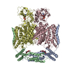
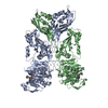
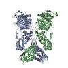
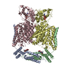
| Entry | Database: PDB / ID: 7jm7 | |||||||||
|---|---|---|---|---|---|---|---|---|---|---|
| Title | Structure of human CLC-7/OSTM1 complex | |||||||||
 Components Components |
| |||||||||
 Keywords Keywords | MEMBRANE PROTEIN / Lysosomal / chloride-proton antiporter / chloride transport / ion transport / proton transport / complex / glycosylated | |||||||||
| Function / homology |  Function and homology information Function and homology informationtransepithelial chloride transport / voltage-gated chloride channel activity / antiporter activity / chloride transmembrane transporter activity / chloride channel activity / chloride channel complex / osteoclast differentiation / Stimuli-sensing channels / lysosomal membrane / intracellular membrane-bounded organelle ...transepithelial chloride transport / voltage-gated chloride channel activity / antiporter activity / chloride transmembrane transporter activity / chloride channel activity / chloride channel complex / osteoclast differentiation / Stimuli-sensing channels / lysosomal membrane / intracellular membrane-bounded organelle / ATP binding / membrane / cytosol Similarity search - Function | |||||||||
| Biological species |  Homo sapiens (human) Homo sapiens (human) | |||||||||
| Method | ELECTRON MICROSCOPY / single particle reconstruction / cryo EM / Resolution: 2.82 Å | |||||||||
 Authors Authors | Schrecker, M. / Hite, R. | |||||||||
| Funding support |  United States, 2items United States, 2items
| |||||||||
 Citation Citation | Journal: Elife / Year: 2020 Title: Cryo-EM structure of the lysosomal chloride-proton exchanger CLC-7 in complex with OSTM1. Authors: Marina Schrecker / Julia Korobenko / Richard K Hite / Abstract: The chloride-proton exchanger CLC-7 plays critical roles in lysosomal homeostasis and bone regeneration and its mutation can lead to osteopetrosis, lysosomal storage disease and neurological ...The chloride-proton exchanger CLC-7 plays critical roles in lysosomal homeostasis and bone regeneration and its mutation can lead to osteopetrosis, lysosomal storage disease and neurological disorders. In lysosomes and the ruffled border of osteoclasts, CLC-7 requires a β-subunit, OSTM1, for stability and activity. Here, we present electron cryomicroscopy structures of CLC-7 in occluded states by itself and in complex with OSTM1, determined at resolutions up to 2.8 Å. In the complex, the luminal surface of CLC-7 is entirely covered by a dimer of the heavily glycosylated and disulfide-bonded OSTM1, which serves to protect CLC-7 from the degradative environment of the lysosomal lumen. OSTM1 binding does not induce large-scale rearrangements of CLC-7, but does have minor effects on the conformation of the ion-conduction pathway, potentially contributing to its regulatory role. These studies provide insights into the role of OSTM1 and serve as a foundation for understanding the mechanisms of CLC-7 regulation. | |||||||||
| History |
|
- Structure visualization
| Movie |
Movie viewer |
|---|---|
| Structure viewer | Molecule: MolmilJmol/JSmol |
- Downloads & links
-Download
| PDBx/mmCIF format | 7jm7.cif.gz | 618.4 KB | Display | PDBx/mmCIF format |
|---|---|---|---|---|
| PDB format | pdb7jm7.ent.gz | 518.3 KB | Display | PDB format |
| PDBx/mmJSON format | 7jm7.json.gz | Tree view | PDBx/mmJSON format | |
| Others |  Other downloads Other downloads |
-Validation report
| Summary document | 7jm7_validation.pdf.gz | 1.5 MB | Display | wwPDB validaton report |
|---|---|---|---|---|
| Full document | 7jm7_full_validation.pdf.gz | 1.5 MB | Display | |
| Data in XML | 7jm7_validation.xml.gz | 56.1 KB | Display | |
| Data in CIF | 7jm7_validation.cif.gz | 82.1 KB | Display | |
| Arichive directory | https://data.pdbj.org/pub/pdb/validation_reports/jm/7jm7ftp://data.pdbj.org/pub/pdb/validation_reports/jm/7jm7 | HTTPS FTP |
-Related structure data
| Related structure data |  22389MC  7jm6C C: citing same article ( M: map data used to model this data |
|---|---|
| Similar structure data |


-Links
 PDBj
PDBj
- Assembly
| Deposited unit | 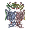
| ||||||||||||||||||
|---|---|---|---|---|---|---|---|---|---|---|---|---|---|---|---|---|---|---|---|
| 1 |
| ||||||||||||||||||
| Noncrystallographic symmetry (NCS) | NCS domain:
NCS ensembles :
|
-Components
-Protein , 2 types, 4 molecules BDAC
| #1: Protein | Mass: 37290.570 Da / Num. of mol.: 2 Source method: isolated from a genetically manipulated source Source: (gene. exp.) Homo sapiens (human) / Gene: OSTM1, GL, HSPC019, UNQ6098/PRO21201 / Production host: Homo sapiens (human) / References: UniProt: Q86WC4#2: Protein | Mass: 88773.875 Da / Num. of mol.: 2 Source method: isolated from a genetically manipulated source Source: (gene. exp.) Homo sapiens (human) / Gene: CLCN7 / Production host: Homo sapiens (human) / References: UniProt: P51798 |
|---|
-Sugars , 3 types, 14 molecules 
| #3: Polysaccharide | Source method: isolated from a genetically manipulated source #4: Polysaccharide | 2-acetamido-2-deoxy-beta-D-glucopyranose-(1-4)-2-acetamido-2-deoxy-beta-D-glucopyranose Source method: isolated from a genetically manipulated source #5: Sugar | ChemComp-NAG /  Type: D-saccharide, beta linking / Mass: 221.208 Da / Num. of mol.: 8 / Source method: obtained synthetically / Formula: C8H15NO6 / Feature type: SUBJECT OF INVESTIGATION Type: D-saccharide, beta linking / Mass: 221.208 Da / Num. of mol.: 8 / Source method: obtained synthetically / Formula: C8H15NO6 / Feature type: SUBJECT OF INVESTIGATION |
|---|
-Non-polymers , 5 types, 48 molecules 


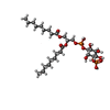

| #6: Chemical |  Mass: 507.181 Da / Num. of mol.: 2 / Source method: obtained synthetically / Formula: C10H16N5O13P3 / Comment: ATP, energy-carrying molecule*YM Mass: 507.181 Da / Num. of mol.: 2 / Source method: obtained synthetically / Formula: C10H16N5O13P3 / Comment: ATP, energy-carrying molecule*YM#7: Chemical |  Mass: 24.305 Da / Num. of mol.: 2 / Source method: obtained synthetically / Formula: Mg Mass: 24.305 Da / Num. of mol.: 2 / Source method: obtained synthetically / Formula: Mg#8: Chemical | ChemComp-CL /  Mass: 35.453 Da / Num. of mol.: 6 / Source method: obtained synthetically / Formula: Cl Mass: 35.453 Da / Num. of mol.: 6 / Source method: obtained synthetically / Formula: Cl#9: Chemical |  Mass: 694.640 Da / Num. of mol.: 2 / Source method: obtained synthetically / Formula: C27H52O16P2 / Feature type: SUBJECT OF INVESTIGATION Mass: 694.640 Da / Num. of mol.: 2 / Source method: obtained synthetically / Formula: C27H52O16P2 / Feature type: SUBJECT OF INVESTIGATION#10: Water | ChemComp-HOH / | Mass: 18.015 Da / Num. of mol.: 36 / Source method: isolated from a natural source / Formula: H2O |
|---|
-Details
| Has ligand of interest | Y |
|---|
-Experimental details
-Experiment
| Experiment | Method: ELECTRON MICROSCOPY |
|---|---|
| EM experiment | Aggregation state: PARTICLE / 3D reconstruction method: single particle reconstruction |
- Sample preparation
| Component | Name: Human CLC-7/OSTM1 complex / Type: COMPLEX / Entity ID: #1-#2 / Source: RECOMBINANT | ||||||||||||||||||||||||
|---|---|---|---|---|---|---|---|---|---|---|---|---|---|---|---|---|---|---|---|---|---|---|---|---|---|
| Molecular weight | Experimental value: NO | ||||||||||||||||||||||||
| Source (natural) | Organism:  Homo (humans) Homo (humans) | ||||||||||||||||||||||||
| Source (recombinant) | Organism: Homo sapiens (human) | ||||||||||||||||||||||||
| Buffer solution | pH: 8 | ||||||||||||||||||||||||
| Buffer component |
| ||||||||||||||||||||||||
| Specimen | Conc.: 2 mg/ml / Embedding applied: NO / Shadowing applied: NO / Staining applied: NO / Vitrification applied: YES | ||||||||||||||||||||||||
| Specimen support | Grid material: GOLD / Grid mesh size: 400 divisions/in. / Grid type: Quantifoil R1.2/1.3 | ||||||||||||||||||||||||
| Vitrification | Instrument: FEI VITROBOT MARK IV / Cryogen name: ETHANE / Humidity: 100 % / Chamber temperature: 277 K |
- Electron microscopy imaging
| Experimental equipment |  Model: Titan Krios / Image courtesy: FEI Company |
|---|---|
| Microscopy | Model: FEI TITAN KRIOS |
| Electron gun | Electron source:  FIELD EMISSION GUN / Accelerating voltage: 300 kV / Illumination mode: FLOOD BEAM FIELD EMISSION GUN / Accelerating voltage: 300 kV / Illumination mode: FLOOD BEAM |
| Electron lens | Mode: OTHER / Nominal magnification: 22500 X / Nominal defocus max: -2700 nm / Nominal defocus min: -1200 nm / Cs: 2.7 mm |
| Image recording | Average exposure time: 8 sec. / Electron dose: 44 e/Å2 / Detector mode: SUPER-RESOLUTION / Film or detector model: GATAN K3 (6k x 4k) |
- Processing
| Software |
| ||||||||||||||||||||||||||||
|---|---|---|---|---|---|---|---|---|---|---|---|---|---|---|---|---|---|---|---|---|---|---|---|---|---|---|---|---|---|
| EM software |
| ||||||||||||||||||||||||||||
| CTF correction | Type: PHASE FLIPPING AND AMPLITUDE CORRECTION | ||||||||||||||||||||||||||||
| Symmetry | Point symmetry: C2 (2 fold cyclic) | ||||||||||||||||||||||||||||
| 3D reconstruction | Resolution: 2.82 Å / Resolution method: FSC 0.143 CUT-OFF / Num. of particles: 327619 / Symmetry type: POINT | ||||||||||||||||||||||||||||
| Atomic model building | Protocol: OTHER / Space: REAL | ||||||||||||||||||||||||||||
| Refinement | Cross valid method: NONE Stereochemistry target values: GeoStd + Monomer Library + CDL v1.2 | ||||||||||||||||||||||||||||
| Displacement parameters | Biso mean: 48.22 Å2 | ||||||||||||||||||||||||||||
| Refine LS restraints |
|
